Session Information
Date: Monday, September 23, 2019
Session Title: Genetics
Session Time: 1:45pm-3:15pm
Location: Les Muses Terrace, Level 3
Objective: Do clinical cognitive profiles and CSF characterisitcs of Abeta1_42, total-tau, phospho-tau and alpha-synuclein during lifetime correspond to histopathological findings post-mortem in LRKK2-associated Parkinson’s disease (PD).
Background: Clinical features of PD patients with LRRK2 mutations (PDLRRK2) resemble those of idiopathic PD (PDidiopathic) with disease progression similar if not somewhat more benign, especially in terms of cognitive decline. Histopathology in PDLRRK2 is variable, including typical Lewy-body pathology with alpha-synuclein aggregation as observed in PDidiopathic but also tau aggregation or nigral degeneration without distinctive histopathology. Among PDLRRK2 the main pathogenic mutation p.G2019S is primarily associated with Lewy body pathology.
Method: Case report of a 64 years old female PD patient carrying the p.G2019S LRRK2-mutation. The patient was regularly assessed over 8 years including Unified Parkinson’s disease Rating Scale part III (UPDRS III), Hoehn and Yahr Scale, Mini Mental Status Examination (MMSE) and Montreal Cognitive Assessment (MoCA). Moreover, repeated CSF-collection (4 timepoints) with measurement of Amyloid-beta1_42, total-Tau, phosphorylated-Tau and total αlpha-synuclein CSF levels was done. After death (21 years of disease duration) brain autopsy with histopathological evaluation was done.
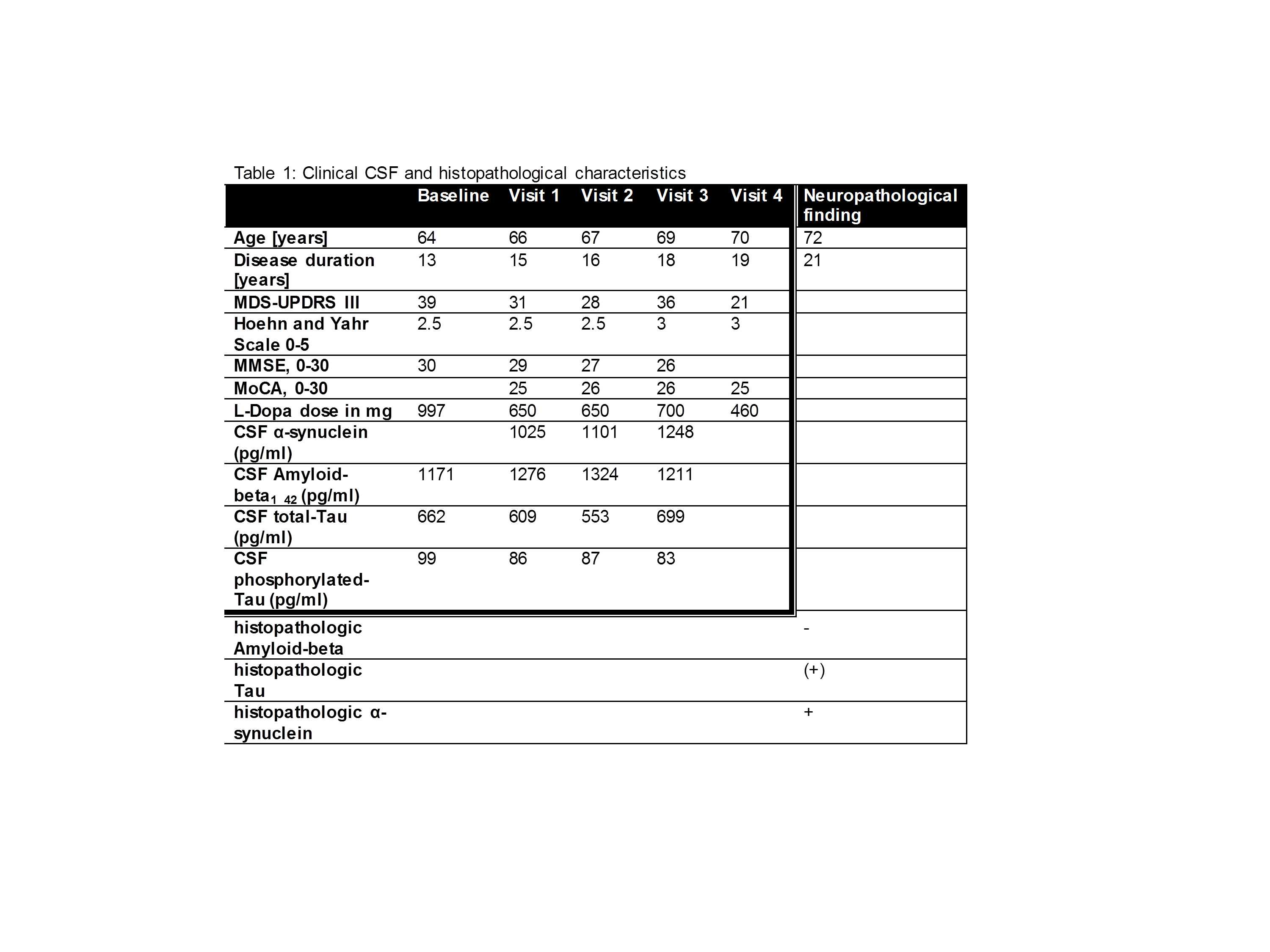
Results: The overall clinical disease course was typical for PDLRRK2 with primarily motor involvement and sparse non-motor manifestation. Of note, cognitive function measured by MMSE and MoCa was preserved and the patient was independent at home until death. CSFprofiles showed normal levels of αlpha-synuclein and of Amyloid-beta1_42 along with slightly elevated levels of total-Tau and phosphorylated-Tau (table 1). Correspondingly, brain autopsy and histopathology showed pronounced Lewy body pathology in substantia nigra pars compacta, locus coeruleus and dorsal ncl. Vagus whereas cortical regions were mostly spared: frontal (Score 0), temporal (Score 1), parietal (Score 0). No senile Amyloid plaques or accumulation in vessel walls were observed. Phospho-tau-aggregation was sparse and only seen in the entorhinal and transentorhinal cortex.
Conclusion: Longitudinally assessed clinical and CSF biomarker profiles of PD patients might be indicative for histopathological pattern. This might help to stratify PD patients according to their primary underlying pathogenesis.
To cite this abstract in AMA style:
I. Wurster, S. Lerche, I. Lachmann, M. Neumann, T. Gasser, K. Brockmann. Longitudinal CSF-profile and brain pathology mirrors clinical course of LRRK2-Related Parkinson’s Disease [abstract]. Mov Disord. 2019; 34 (suppl 2). https://www.mdsabstracts.org/abstract/longitudinal-csf-profile-and-brain-pathology-mirrors-clinical-course-of-lrrk2-related-parkinsons-disease/. Accessed April 20, 2025.« Back to 2019 International Congress
MDS Abstracts - https://www.mdsabstracts.org/abstract/longitudinal-csf-profile-and-brain-pathology-mirrors-clinical-course-of-lrrk2-related-parkinsons-disease/

