Category: Parkinson's Disease: Neuroimaging
Objective: To elucidate longitudinal brain atrophies in PD with or without RBD and anosmia at baseline.
Background: REM sleep behavior disorder (RBD) and olfactory dysfunction (OD) in early Parkinson’s disease are associated with an increased risk of cognitive decline, which might be related to a different pattern or dynamics of progression of brain pathology in affected individuals. The impact of these two common early features on the trajectory of longitudinal brain volume remains unclear.
Method: This study included the longitudinal MRI and clinical data of 136 de novo PD and 49 healthy controls (HC) provided by the Parkinson’s Progression Markers Initiative (PPMI). PD patients were divided into four groups: those with or without probable RBD (RBD+, RBDSQ cutoff = 5 but 12 patients with score 5 were excluded) and OD+ (anosmia cutoff of UPSIT score = 18). Linear mixed effect models were used to investigate differences in longitudinal cortical atrophic changes in PD with or without RBD or OD, comparing the volume decline to HC.
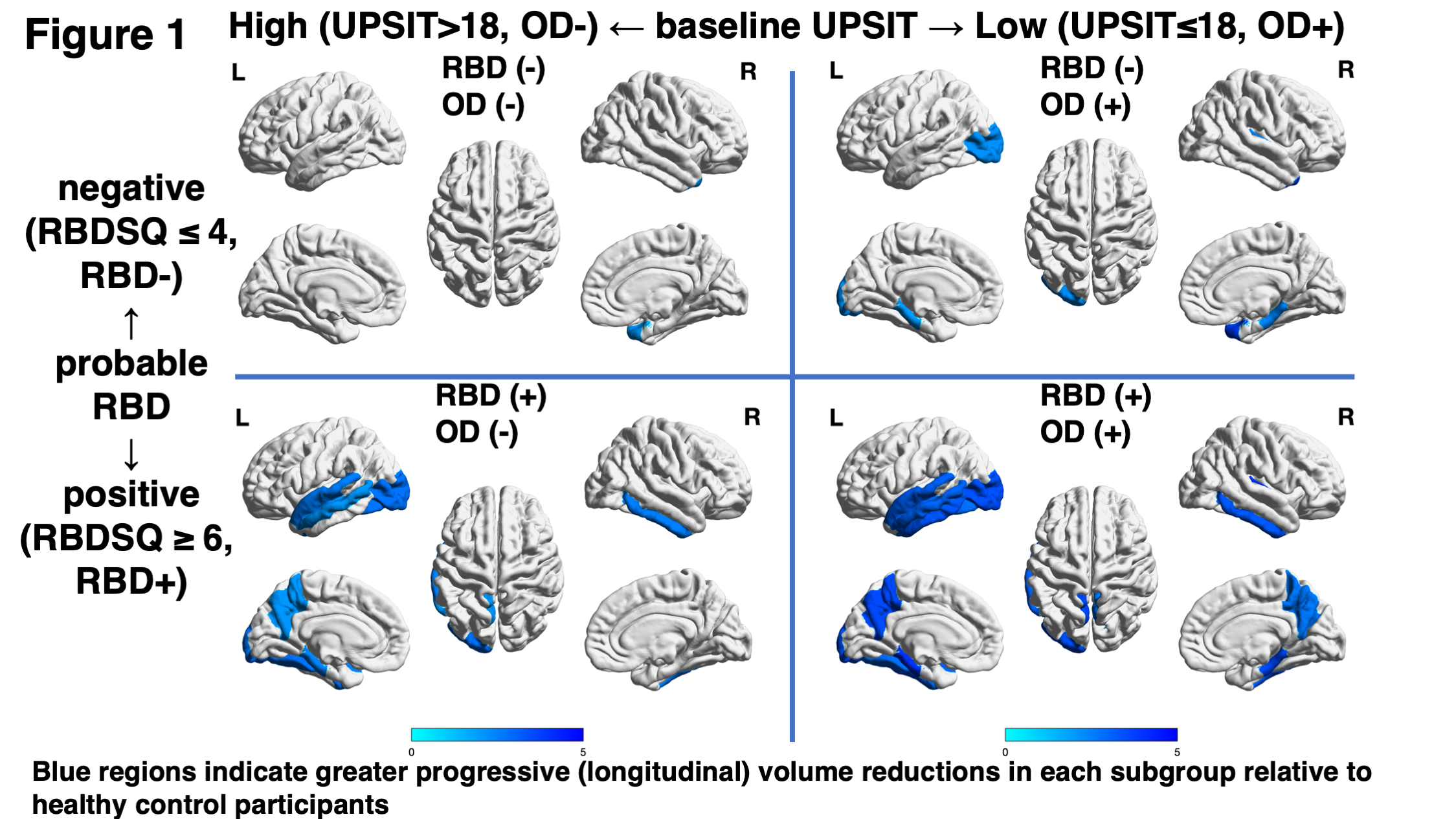
Results: Baseline demographics showed that SCOPA-AUT and MDS-UPDRS part I scores were higher in the RBD+ groups. PD with RBD+/OD+ had higher neurofilament light chain level (NfL). The RBD+/OD+ group (n=19) group had more extensive brain volume decline in the temporal and parietal lobes than others [figure1]. Specifically, this group showed greater atrophic changes in temporal (left fusiform, bilateral inferior temporal, left middle temporal, bilateral parahippocampal, left superior temporal, and right transverse temporal gyri), parietal (bilateral precuneus), and occipital (left lateral occipital cortex) regions. RBD+/OD- (n=16) had volume decline in the left fusiform, left middle temporal, left parahippocampal, left superior temporal, right inferior temporal gyri, and left precuneus. RBD-/OD+ (n=23) showed longitudinal volume reductions in the bilateral parahippocampal, right transverse temporal, left lateral occipital gyri, and right temporal pole. RBD-/OD- (n=66) had a volume decline only in the right temporal pole.
Conclusion: De novo PD patients with pRBD and OD had progressive cortical atrophy in parietal and temporal regions that was more extensive than in patients without these symptoms and those with only one of these features. The affected regions are critical for cognitive function and their greater involvement might underly an increased risk of cognitive decline in PD patients with RBD and OD.
To cite this abstract in AMA style:
K. Kawabata, E. Bagarinao, K. Seppi, W. Poewe. Longitudinal brain volume decline in PD with RBD and olfactory dysfunction [abstract]. Mov Disord. 2023; 38 (suppl 1). https://www.mdsabstracts.org/abstract/longitudinal-brain-volume-decline-in-pd-with-rbd-and-olfactory-dysfunction/. Accessed December 15, 2025.« Back to 2023 International Congress
MDS Abstracts - https://www.mdsabstracts.org/abstract/longitudinal-brain-volume-decline-in-pd-with-rbd-and-olfactory-dysfunction/

