Session Information
Date: Wednesday, September 25, 2019
Session Title: Neuroimaging
Session Time: 1:15pm-2:45pm
Location: Les Muses Terrace, Level 3
Objective: To assess longitudinal abnormlities throughout the corticospinal tract in Parkinson’s Disease patients.
Background: Parkinson’s Disease (PD) is a neurodegenerative progressive disease might become a pandemic disease in the recent future[1] [2]. Considering that, the pathophysiology is still not fully understood, the longitudinal analysis allows an in vivo observation of brain alterations over time, making it possible to infer some hypothesis about disease progression.
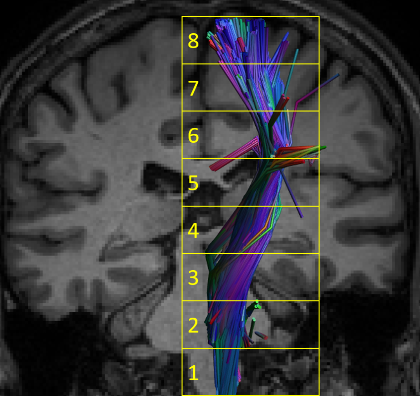
Method: DTI microstructural parameters of the cortical-spinal tract (CST) were obtained using the software ExploreDTI. The DTI images were corrected for signal drift, Gibbs’ rings, susceptibility artifact, Eddy currents and movement and then registered to a subject T1 weighted image. We used a semi-automatic methodology for fiber tracking based on strategies drawn on a reference template. The tracts were uniformly resampled and divided into 8 segments [Figure1]. Thus, we analyzed average FA (FA_avg), AD (AD_avg) and RD (RD_avg) values of right and left CST to avoid type I error. We performed a one-way repeated-measures analysis of covariance. However, since the time between acquisitions varied greatly 1-6 years), we included this interval as a nuisance covariate. Three models were built, one for each measure.
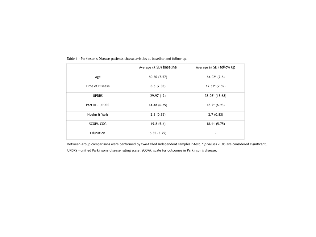
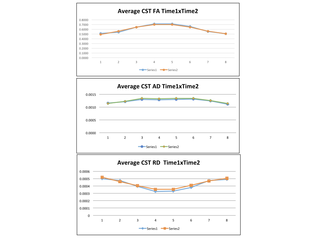
Results: Patients demographics are described in table 1. [Table1]. Overall, we found a significant multivariate effect of time in FA_avg (F8,31= 3.85, p = 0.003) and RD_avg (F8,31= 46.55, p < 0.001) but not AD_avg (F8,31= 1.54, p = 0.181). Univariate post-hoc pairwise comparisons showed a significant reduction of FA over time in segments 1 (p = 0.016) and 4 (p = 0.042) of the CST. For RD_avg, we found increased values over time in segments 4 (p = 0.003), 5 (p = 0.016) and 6 (p = 0.003). However, RD_avg were decreased at baseline compared to the follow-up acquisition (p < 0.001).However, ADM were decreased at baseline compared to the follow-up acquisition in all, but the first segment, in a univariate post-hoc comparison.
Conclusion: We found an increase in FA and a decrease in RD values, indicating neurodegeneration in the CST in PD patients in different segments. These diffusion measures might be a valuable biomarker of disease progression. Larger longitudinal studies are needed to indicate if these values can be used as sensitive and clinically meaningful measures of disease progression in PD.
References: 1. Tysnes, O.B. and A. Storstein, Epidemiology of Parkinson’s disease. J Neural Transm (Vienna), 2017. 124(8): p. 901-905. 2. Dorsey, E.R. and B.R. Bloem, The Parkinson Pandemic-A Call to Action. JAMA Neurol, 2018. 75(1): p. 9-10.
To cite this abstract in AMA style:
R. Guimarães, L. Piovesana, P. Azevedo, B. Campos, F. Cendes. Longitudinal Analysis of the Corticospinal tract microstructure in Parkinson’s Disease Patients [abstract]. Mov Disord. 2019; 34 (suppl 2). https://www.mdsabstracts.org/abstract/longitudinal-analysis-of-the-corticospinal-tract-microstructure-in-parkinsons-disease-patients/. Accessed April 21, 2025.« Back to 2019 International Congress
MDS Abstracts - https://www.mdsabstracts.org/abstract/longitudinal-analysis-of-the-corticospinal-tract-microstructure-in-parkinsons-disease-patients/


