Category: Parkinson's Disease: Neuroimaging
Objective: To assess longitudinal diffusion tensor imaging differences between PD patients in 8 corticospinal tract segments.
Background: Parkinson’s disease (PD) is clinically defined by the presence of cardinal motor symptoms such as: rest tremor, rigidity and bradicinesia. The corticospinal tract is the major motor tract and is related to a variety of movements, such as gait, writing, typing, buttoning, which are all impaired in PD.
Method: We included 39 PD patients (mean age 60 years at t1 and 63 at t2). Demographics are described in table 1.
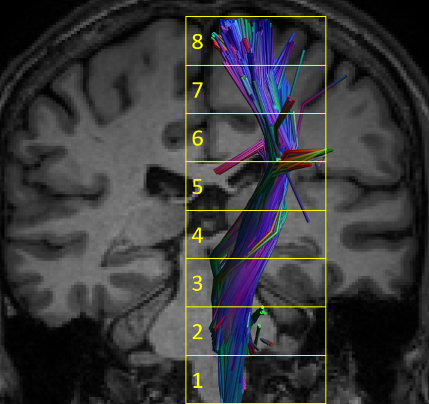
DTI microstructural parameters of the cortical-spinal tract (CST) were obtained using the software ExploreDTI. We used a semi-automatic methodology for fiber tracking based on strategies drawn on a reference template. The tracts were uniformly resampled and divided into 8 segments [Figure1]. Statistical analysis: First, we tested whether there were asymmetries between right and left CST segments using paired t-tests for each acquisition time (see Results). Since CST is a midline tract and we did not find any significant differences between right and left CST, we averaged right and left CST values for FA (FAm), AD (ADm) and RD (RDm). This strategy simplifies the analysis and minimizes type I error by reducing the number of variables.The longitudinal model, with PD patients only, included time of scan (scan-1 and scan-2), segment (two to seven) and a time of scan-by-segment interaction. We also included disease duration as a covariate. The models were best fitted with first-order autoregressive matrix as variance-covariance structure, using log as link function to gamma-regression distribution (to adjust for right skewness in the data). We evaluated the best fitting for the final models by using the Akaike Information Criteria. All the pairwise comparisons in the models were corrected using the Holm-Sidaq procedure. We accepted p < 0.05 as statistically significant.
Results: Diffusion metrics changes – PD group over time – Significant effect of CST segments but no significant effect of time nor interaction. No significant changes in none of the segments between scan-1 and scan-2.
Conclusion: There is no difference in DTI measures along time in the CST in PD patients. However, the is a wide variety of FA measures along the tract, suggesting that there might be a more an ascending compromise of the tract, as previously suggested by other authors.
To cite this abstract in AMA style:
R. Guimaraes, L. Ramalho, B. Campos, F. Cendes. Longitudinal Analysis of the Corticospinal Tract in Parkinson’s disease Patients [abstract]. Mov Disord. 2022; 37 (suppl 2). https://www.mdsabstracts.org/abstract/longitudinal-analysis-of-the-corticospinal-tract-in-parkinsons-disease-patients/. Accessed April 18, 2025.« Back to 2022 International Congress
MDS Abstracts - https://www.mdsabstracts.org/abstract/longitudinal-analysis-of-the-corticospinal-tract-in-parkinsons-disease-patients/


![Figure [2].001](https://www.mdsabstracts.org/wp-content/uploads/2022/09/0650_0914_001824_002.jpeg)
![table [1].001](https://www.mdsabstracts.org/wp-content/uploads/2022/09/0650_0914_001824_003.jpeg)