Objective: The aim of this study was to test the utility of isolating L1 Cell Adhesion Molecule (L1CAM)-derived extracellular vesicles (EVs) in quantification of glucose metabolism markers and the assessment of potential indicators of brain insulin resistance in persons with Parkinson’s disease (PwPD) and healthy controls (HCs).
Background: Similar molecular processes have been identified in the development of PD and type 2 diabetes mellitus (T2DM) [1]. Insulin resistance is a central feature of T2DM pathology, and recent reports provide evidence for its occurrence in the brain and contribution in mechanisms of neurodegeneration [2]. Glucagon-like peptide 1(GLP-1) analogues are drugs that promote insulin signalling, approved for the treatment of T2DM, and under intensive research efforts to assess their efficacy in slowing disease progression in PD. No specific, easily accessible biomarker exists to measure insulin sensitivity in the brain, which would be valuable both in patient selection for targeted treatments as well as a measure of treatment effect.
Method: EVs were isolated in plasma of 78 PwPD and 24 HCs according to previously described method [3]. The method was evaluated with three rounds of SDS-PAGE and Western blot. Neuronal enrichment was performed with immune capture for neuronal surface antigen L1CAM/CD171. Mesoscale multiplex metabolic panel was used for the measurement of insulin, c-peptide, pancreatic polypeptide, GLP-1, gastric inhibitory polypeptide, leptin and glucagon. ELISA was used to measure phosphorylated forms of insulin receptor substrate 1 (IRS1).
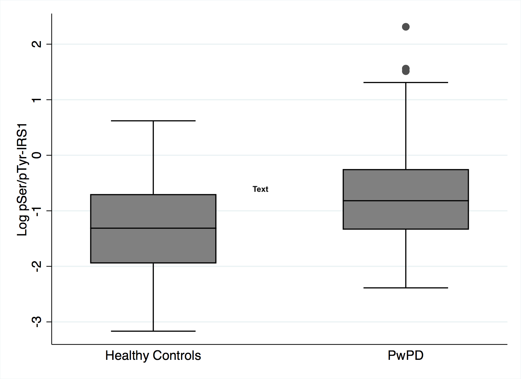
Results: Peripheral fasting insulin levels, glucose levels and HOMA index did not differ between PwPD and HCs (p=0.8; p=0.4 and p=0.9 respectively). L1CAM-exosome levels of insulin did not differ between PwPD and HCs, whereas the ratio of pSer-IRS1/pTyr-IRS1 was higher in PwPD compared to HCs (Fig.1; p=0.04), indicating decreased insulin sensitivity in the PD group.
Conclusion: Our findings show that although peripheral insulin sensitivity does not differ between PwPD and HCs, there are signs of decreased insulin sensitivity in the receptor level, in the brain of PwPD, as assessed by the ratio of pSer/pTyr-IRS1 in L1CAM-derived exosomes. Further studies are necessary to confirm the robustness of plasma isolated EVs as easily accessible carriers of real-time information about brain dysfunction.
References: [1] JA Santiago and JA Potashkin, Trends Mol Med 19 (3), 176 (2013).
[2] SE Arnold et al., Nat Rev Neurol 14 (3), 168 (2018).
[3] D Kapogiannis et al., FASEB J 29 (2), 589 (2015).
To cite this abstract in AMA style:
I. Markaki, W. Paslawski, T. Ntetsika, P. Svenningsson. Isolation of L1CAM-extracellular vesicles reveals signs of insulin resistance in Parkinson’s disease [abstract]. Mov Disord. 2023; 38 (suppl 1). https://www.mdsabstracts.org/abstract/isolation-of-l1cam-extracellular-vesicles-reveals-signs-of-insulin-resistance-in-parkinsons-disease/. Accessed April 20, 2025.« Back to 2023 International Congress
MDS Abstracts - https://www.mdsabstracts.org/abstract/isolation-of-l1cam-extracellular-vesicles-reveals-signs-of-insulin-resistance-in-parkinsons-disease/

