Category: Parkinson's Disease: Neuroimaging
Objective: The aim of the study was to determine a technical parameter of foveal pathology that is best suited for discriminating Parkinson’s disease (PD) eyes from those of healthy controls and to assess correlations between impaired contrast sensitivity and foveal shape in PD patients.
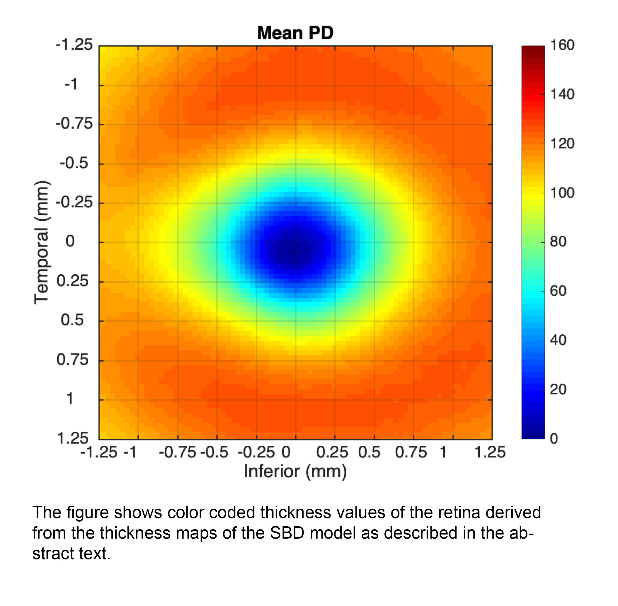
Background: The visual system is affected in PD. PD patients demonstrate loss in spatiotemporal contrast sensitivity (CS), which parallels with motor fluctuations. Foveal remodeling that specifies the Symmetry, Depth and Breadth of the fovea (SBD model) is a promising tool to qualify and quantify retinal pathology in PD.
Method: We characterized the fovea in 48 Parkinson’s disease patients and 45 control subjects by optical coherence tomography (OCT) to examine the intrinsic model of foveal configuration. Additionally the following demographic and clinical data were collected: Age, gender, disease duration, Hoehn & Yahr stage, medication at time of examination, the Unified Parkinson’s Disease Rating Scale, Part III (UPDRS-III), the Montreal Cognitive Assessment (MoCA) and the Pelli-Robson Contrast Sensitivity Test.
Results: In Principal Component Analysis the best combination of parameters of the eight coefficients of the SBD model was A21 A22 with an AUC of 60.03%. CS examination showed a highly significant difference in CS between PD and controls. There was a significant correlation between binocular CS and the A0 (p=0.013) and A22 (p=0.043) SBD model parameters.
Conclusion: The examined model quantifies structural changes in the fovea of Parkinson’s disease patients that are correlated with a decline in contrast sensitivity. We could show that there is a significant correlation between functional vision deficits and foveal pit architecture changes in PD as shown by the SBD model. It is unclear however, whether changes of the foveal slope configuration are associated with motor decline or cognitive decline in PD.
References: Spund B, Ding Y, Liu T, Selesnick I, Glazman S, Shrier EM, et al. Remodeling of the fovea in Parkinson disease. J Neural Transm. 2012. Available: http://link.springer.com/article/10.1007/s00702-012-0909-5/fulltext.html Ding Y, Spund B, Glazman S, Shrier EM, Miri S, Selesnick I, et al. Application of an OCT data-based mathematical model of the foveal pit in Parkinson disease. J Neural Transm. 2014;121: 1367–1376. Available: http://link.springer.com/10.1007/s00702-014-1214-2
To cite this abstract in AMA style:
E. Pinkhardt, A. Ding, S. Slotnick, J. Kassubek, A. Ludolph, S. Glazman, I. Selesnick, I. Bodis-Wollner. Intrinsic Remodeling of the Fovea is Correlated with Contrast Sensitivity Loss in Parkinson’s Disease [abstract]. Mov Disord. 2020; 35 (suppl 1). https://www.mdsabstracts.org/abstract/intrinsic-remodeling-of-the-fovea-is-correlated-with-contrast-sensitivity-loss-in-parkinsons-disease/. Accessed April 21, 2025.« Back to MDS Virtual Congress 2020
MDS Abstracts - https://www.mdsabstracts.org/abstract/intrinsic-remodeling-of-the-fovea-is-correlated-with-contrast-sensitivity-loss-in-parkinsons-disease/

