Category: Surgical Therapy: Parkinson's Disease
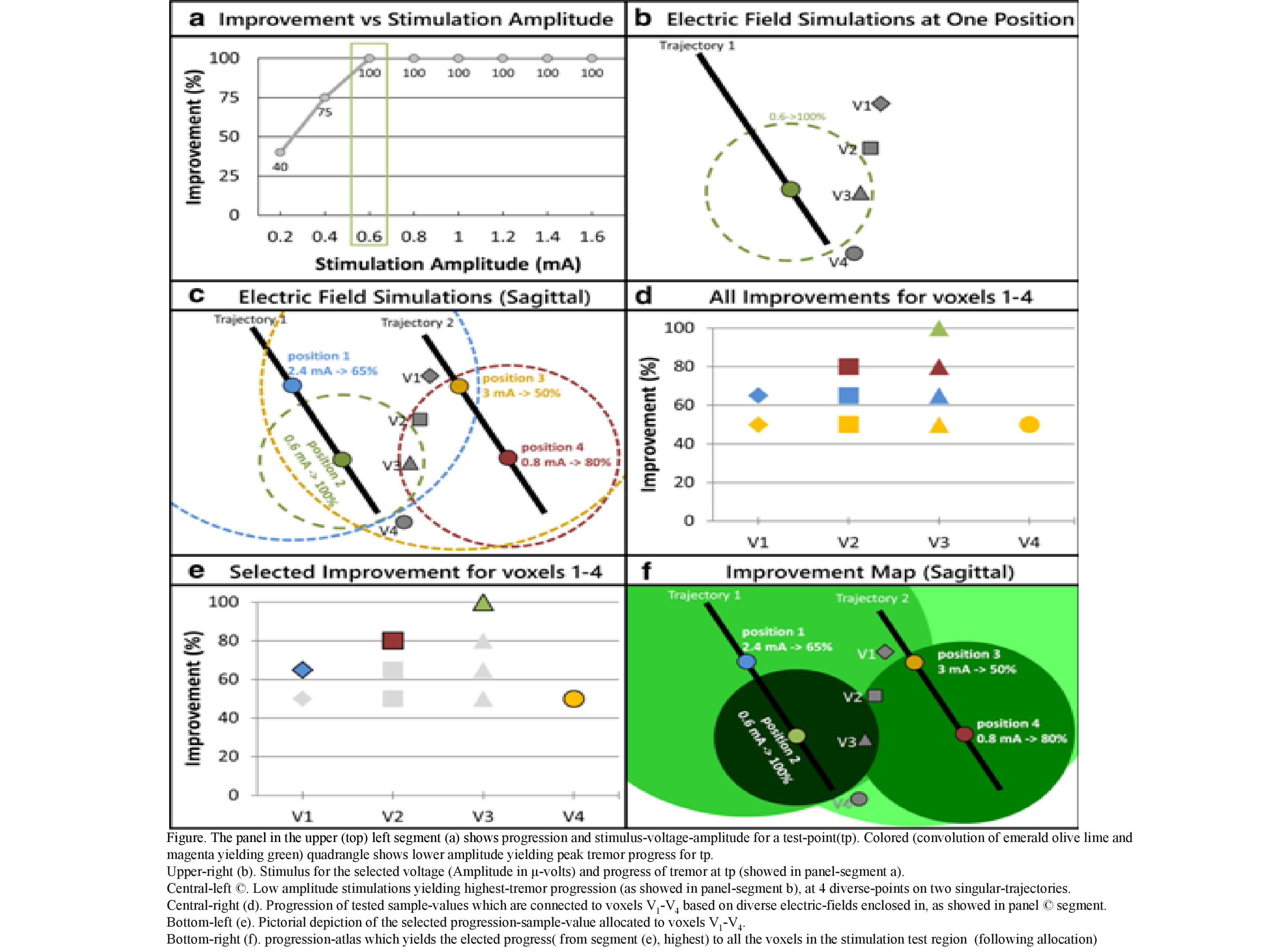
Objective: To convolve the subject-specific (SS) electric-stimulus-field (ESF) with tremor-progress and progression quantified through accelerometer and stimulus-induced adverse side-effects (SIASE), exhibit visually (by eye) in the form of stimulus-atlases superimposed on the subject-specific-anatomy (SSA) and to aid in surgical-operation team works (for decision making, i.e., choosing the optimal-implant-site (OIS) for the continual stimulus DBS electrode.
Background: DBS is a well established surgical—method in MDs especially for advanced idiopathic Parkinson’s disease. The stimulus electrode point in Parkinson’s disease brain is a key for successful management. Wide assessments of perfection and adverse dyskinesias (side-effects) of stimulus on singular points especially for diverse amplitudes are achieved. Yet, the information has to be examined carefully for choosing the optimal site of the electrode. This study introduces a novel stimulus-atlases procedure that outlines and mentally pictures’ (or visually by eye) the massive data gathered which helps in discovering the optimal electrode site. Study carried at tertiary hospital South India.
Method: A novel technique called “Stimulus-Atlases” introduced in this retrospective study, which precise and pictures (by eye) the immense data amid the goal to aid in spotting the best and effective lead-point. It convolves 3 ways: sketch of the pertinent anatomical-structures, evaluation of cardinal motor symptoms of PD, and subject-specific (SS) electric-field-models(EFMs). By these ways, each voxel in the stimulus-area is allotted a value of sign and symptom-progress, yielding the dissection of stimulus-area into regions by singular progress-levels. The method was applied to 12 PD subjects at a tertiary care university hospital and research center in south India.
Results: Results showed that the highly advanced area is often in the STN posterior area only. The pictorial representation of stimulus-atlases can be viewed in Figure.
This retrospective study utility tool of the stimulus atlases is very useful in discovering the optimal electrode site to implant in a PD-patient prognostically.
Conclusion: The procedure explained in this study has the likely latent to condense the DBS operation team-work in discovering perfect insertion site of the electrode and to aid and accelerate the use of course-electrodes.
References: 1. Herrington TM, Cheng JJ, Eskandar EN (2016) Mechanisms of deep brain stimulation. J Neurophysiol 115(1):19–38. https://doi. org/10.1152/jn.00281.2015 2. Abosch A, Timmermann L, Bartley S, Rietkerk HG, Whiting D, Connolly PJ, Lanctin D, Hariz MI (2013) An international survey of deep brain stimulation procedural steps. Stereotact Funct Neurosurg 91(1):1–11. https://doi.org/10.1159/000343207 3. Hemm S, Wårdell K (2010) Stereotactic implantation of deep brain stimulation electrodes: a review of technical systems, methods and emerging tools. Med Biol Eng Comput 48(7):611–624. https://doi. org/10.1007/s11517-010-0633-y 4. V. Rama Raju, et.al., “The Role of Microelectrode Recording (MER) in STN DBS Electrode Implantation”, IFMBE Proceedings, Springer Nature, Vol. 51, World Congress on Medical Physics and Biomedical Engineering, June 7-12, 2015, Toronto, Canada, Pp: 1204-1208, www.wc2015.org
To cite this abstract in AMA style:
V. Rama Raju, L. Neerati, A. Dabbeta, H. Yeruva, S. Konda, N. Guguloth. Intraoperative test objectively for discovering optimal electrode site for effective imaging (by eye) and bilateral STN-DBS stereotactic functional neurosurgery [abstract]. Mov Disord. 2020; 35 (suppl 1). https://www.mdsabstracts.org/abstract/intraoperative-test-objectively-for-discovering-optimal-electrode-site-for-effective-imaging-by-eye-and-bilateral-stn-dbs-stereotactic-functional-neurosurgery/. Accessed April 21, 2025.« Back to MDS Virtual Congress 2020
MDS Abstracts - https://www.mdsabstracts.org/abstract/intraoperative-test-objectively-for-discovering-optimal-electrode-site-for-effective-imaging-by-eye-and-bilateral-stn-dbs-stereotactic-functional-neurosurgery/

