Category: Parkinson's Disease: Neuroimaging
Objective: This study is aimed to evaluate the longitudinal associations between cardiac sympathetic denervation and cognitive status in Parkinson’s disease (PD).
Background: There lacks an appropriate extracranial biomarker that delineates endophenotypes of PD at early stage and reflects the neurodegenerative process across time. Evaluation of myocardial sympathetic nerve terminals could be a good candidate.
Method: One hundred and twenty-two early Parkinson’s disease patients were recruited in this longitudinal study. All were examined with positron emission tomography (PET) using 18F-N-(3-fluoropropyl)-2beta-carbon ethoxy-3beta-(4-iodophenyl) nortropane (18F-FP-CIT) and 123I-meta-iodobenzylguanidine myocardial scintigraphy. Cardiac scans were re-examined two or three times. Patients were sub-grouped into the sympathetic denervated group, those without evidence of denervated myocardium in the first and subsequent scans, and the converters whose myocardium was found to be impaired in the subsequent scans. Ninety-nine patients were initially assessed of their cognition with comprehensive neuropsychological tests, and forty-six were re-assessed. Any associations between cardiac denervation subtypes and presynaptic dopamine transporter densities were investigated. The cognitive status across the subgroups was explored and studied how it altered over disease progression in relevance to the changes of cardiac sympathetic denervation.
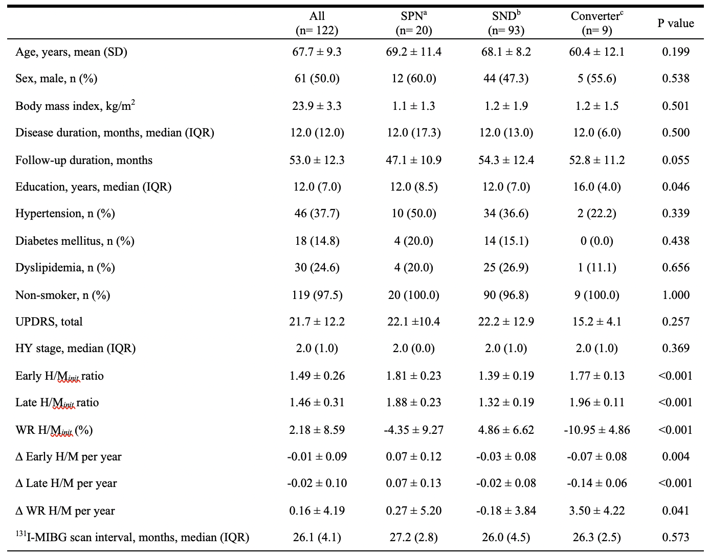
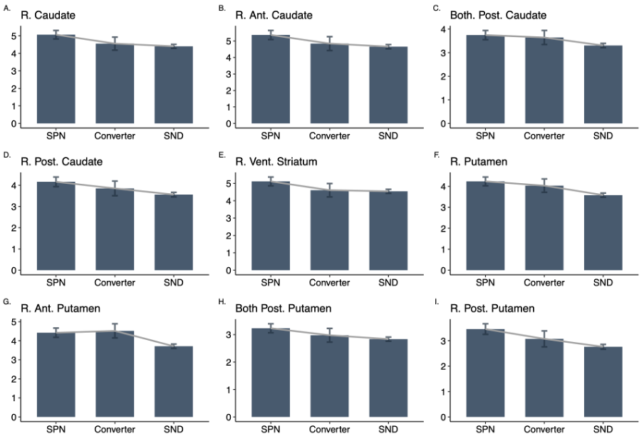
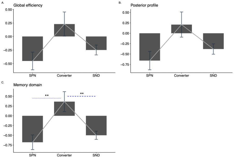
Results: Early PD with mild parkinsonism about 4-5 years of follow-ups were enrolled. The mean age was 67.7 ± 9.3 years, and disease duration was 12.0 (interquartile range, IQR, 12.0) months. Total UPDRS was 21.7 ± 12.2. Cross-sectional comparisons of presynaptic monoamine transporter availability with pre-defined order of cardiac denervation groups revealed the parallel degeneration.[figure1] A quadratic correlation between cardiac catecholamine capacity with cognition was observed at initial and its associations faded with time.[figure2] Its association and disappearance across the disease progression was interpreted to reflect the early neurobiology of PD.
Conclusion: Extracranial cardiac catecholaminergic gradient was observed and it was assumed to mirror the central compensatory neurobiology in early PD.
To cite this abstract in AMA style:
S-W. Yoo, J-S. Kim. Intermediate cardiac denervation in early Parkinson’s disease [abstract]. Mov Disord. 2022; 37 (suppl 2). https://www.mdsabstracts.org/abstract/intermediate-cardiac-denervation-in-early-parkinsons-disease/. Accessed April 18, 2025.« Back to 2022 International Congress
MDS Abstracts - https://www.mdsabstracts.org/abstract/intermediate-cardiac-denervation-in-early-parkinsons-disease/


