Category: Parkinson's Disease: Cognitive functions
Objective: To examine whether PD-MCI patients have increased plasma neurofilament light chain (NfL) levels compared to PD with normal cognition (PD-NC) and healthy controls (HC).
Background: Diagnosis of mild cognitive impairment in Parkinson’s disease (PD-MCI) is currently based on the application of MDS criteria or the use of cut-off values on validated neuropsychological scales. Heterogeneity on the tests used by different research groups or centers limits the generalization of results, making it difficult to establish firm conclusions on the neural bases of PD-MCI, or to determine at the single-patient level which patients are at higher risk of progressive cognitive deterioration.
Neurofilament light chain (NfL) blood levels is an accepted marker of neuroaxonal damage, and it has been shown to act as a marker of PD severity and progression1.
Method: Cross-sectional study including 58 non-demented PD patients (32 PD-MCI, 26 PD-NC), and 26 HC. Plasma NfL was measured with the Simoa Human NF-light Advantage kit using the Single Molecule Array technology.
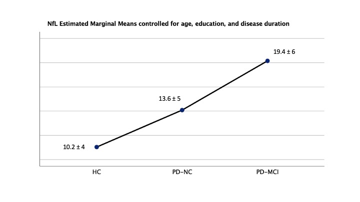
Results: PD-MCI patients were significantly older (p<0.01) and less educated (p=0.042) than PD-NC, but did not differ in gender, disease duration and dopaminergic doses. Univariate ANCOVA analysis controlling for age, education, and evolution showed significantly higher plasma NfL levels in the PD-MCI group than in PD-NC and HC (19.4 ± 6 vs. 13.6 ± 5, and 10.2 ± 4 pg/mL, respectively, F=12.6; p <0.001). Bonferroni post-hoc comparisons showed NfL levels to significantly differentiate PD-MCI and PD-NC patients (I-J -4.07; p<0-01), and both PD-MCI (p<0.001) and PD-NC groups (p=0.03) from HC.
Higher NfL levels correlated significantly with measures of global cognitive function (PD-CRS r=-0.39, p=0.002; MoCA r=-0.27, p=0.04), verbal fluencies (r=-0.41, p=0.001), and delayed verbal (r=-0.36, p=0.007) and visual memory (r=-0.35, p=0.007).
ROC curve analysis determined NfL levels = 17 pg/ml as the optimal cutoff score for the screening of PD-MCI in our sample (AUC 0.77; sensitivity 70.1%, specificity 75%).
Conclusion: The measurement of plasma NfL levels objectively and accurately differentiates PD patients with and without mild cognitive impairment, and correlates with global cognitive function, verbal fluencies and delayed memory.
References: 1. Lin, C.-H. et al. Blood NfL: a biomarker for disease severity and progression in Parkinson disease. Neurology. https://doi.org/10.1212/WNL.0000000000008088 (2019)
To cite this abstract in AMA style:
J. Pagonabarraga, R. Perez-Gonzalez, H. Bejr-Kasem, J. Marin-Lahoz, A. Horta-Barba, I. Aracil-Bolaños, F. Sampedro, S. Martinez-Horta, J. Perez-Perez, B. Pascual-Sedano, J. Kulisevsky. Increased neurofilament light chain plasma levels as a biological marker of mild cognitive impairment in Parkinson’s disease [abstract]. Mov Disord. 2020; 35 (suppl 1). https://www.mdsabstracts.org/abstract/increased-neurofilament-light-chain-plasma-levels-as-a-biological-marker-of-mild-cognitive-impairment-in-parkinsons-disease/. Accessed January 19, 2026.« Back to MDS Virtual Congress 2020
MDS Abstracts - https://www.mdsabstracts.org/abstract/increased-neurofilament-light-chain-plasma-levels-as-a-biological-marker-of-mild-cognitive-impairment-in-parkinsons-disease/

