Category: Neuroimaging (Non-PD)
Objective: To assess grey matter (GM) volume derived brain-aging in Wilson Disease (WD) using a previously proposed machine learning (ML) algorithm.
Background: GM atrophy is a common magnetic resonance imaging (MRI) finding in WD [1,2], which recently was proposed to be accelerated in patients with neurological WD (neuro-WD), in contrast to those with hepatic manifestations (hep-WD) [3]. However, most studies lack whole-brain volumetric quantification due to the high expenditure of time, resources, and expertise. To overcome this limitation, we use a ML-based workflow to efficiently assess brain-aging in WD based on voxel-wise GM volumes and investigate correlations with functional measures.
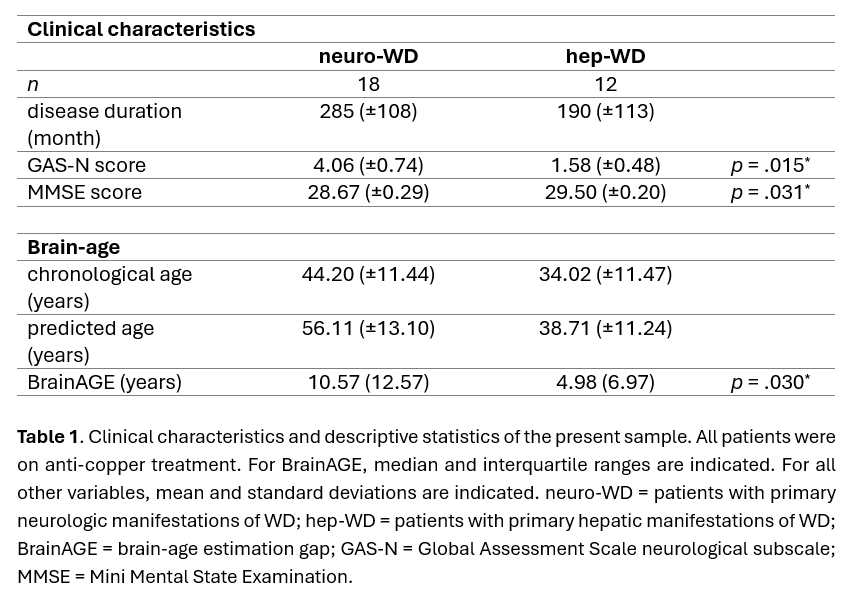
Method: 30 WD patients were scored on the Global Assessment Scale for WD neurological subscale (GAS-N) and the Mini Mental State Examination (MMSE) (see Table 1). 3T MRI was performed according to a protocol adapted from the Lifespan Human Connectome Project in Aging [4]. For brain-age prediction, a Gaussian process regression ML algorithm trained with features derived from voxel-wise GM volumes of large normative neuroimaging datasets [5; https://github.com/juaml/brainage_estimation] was applied to the acquired T1w MR images. The brain-age gap estimate (BrainAGE) score was calculated as the difference between chronological and estimated brain-age.
Results: Within both patient groups, predicted brain-ages were significantly higher than the corresponding chronological ages, neuro-WD: t(17)=-4.83, p<.001, d=1.137; hep-WD: t(11)=-2.93, p=.014, d=0.847. The BrainAGE scores were significantly higher in the neuro-WD than in the hep-WD group, Z=-2.159, p=.030, r=-.394. Overall, higher BrainAGE scores were significantly correlated with higher GAS-N (r=.374, p=.042) and lower MMSE (r=-.364, p=.048) scores.
Conclusion: Our results indicate that brain-aging is significantly accelerated in neuro-WD compared to hep-WD, as supported by evidence of increased atrophy rates in neuro-WD [3]. Furthermore, accelerated brain-aging correlated with higher neurological and cognitive impairment and may thus be a useful biomarker for disease severity. BrainAGE scores for hep-WD were in range of the workflows mean absolute error [5] and should be contrasted to healthy controls in further research. All in all, automated brain-age prediction seems to be a promising tool to simplify the assessment of neurodegeneration and functional decline in patients with WD.
Clinical Characteristics of WD Patients
References: [1] Dusek, P., Smolinski, L., Redzia-Ogrodnik, B., Golebiowski, M., Skowronska, M., Poujois, A., Laurencin, C., Jastrzebska-Kurkowska, I., Litwin, T., & Czlonkowska, A. (2020). Semiquantitative scale for assessing brain MRI abnormalities in Wilson disease: A validation study. Movement Disorders, 35(6), 994-1001. DOI: 10.1002/mds.28018
[2] Viveiros, A., Beliveau, V., Panzer, M., Schaefer, B., Glodny, B., Henninger, B., Tilg, H., Zoller, H., & Scherfler, C. (2021). Neurodegeneration in hepatic and neurologic Wilson’s disease. Hepatology, 74, 1117-1120. DOI: 10.1002/hep.31681
[3] Smolinski, L., Ziemssen, T., Akgun, K., Antos, A., Skowronska, M., Kurkowska-Jastrzebska, I., Czlonkowska, A., & Litwin, T. (2022). Brain atrophy is substantially accelerated in neurological Wilson’s disease: A longitudinal study. Movement Disorders, 37(12), 2446-2451. DOI: 10.1002/mds.29239
[4] Bookheimer, S. Y., Salat, D. H., Terpstra, M., Ances, B. M., Barch, D. M., Buckner, R. L., Burgess, G. C., Curtiss, S. W., Diaz-Santos, M., Elam, J. S., Fischl, B., Greve, D. N., Hagy, H. A., Harms, M. P., Hatch, O. M., Hedden, T., Hodge, C., Japardi, K. C., Kuhn, T. P., Ly, T. K., Smith, S. M., Somerville, L. H., Ugurbil, K., van der Kouwe, A., van Essen, D., Woods, R. P., & Yacoub, E. (2019). The Lifespan Human Connectome Project in Aging: An overview. NeuroImage, 15(185), 335-348. DOI: 10.1016/j.neuroimage.2018.10.009
[5] More, S., Antonopoulos, G., Hoffstaedter, F., Caspers, J., Eickhoff, S. B., & Patil, K. R., for the Alzheimer’s Disease Neuroimaging Initiative (2023). Brain-age predicition: A systematic comparison of machine learning workflows. NeuroImage, 270, 119947. DOI: 10.1016/j.neuroimage.2023.119947
To cite this abstract in AMA style:
A. Hausmann, J. Caspers, S. Kannenberg, S. More, K. Patil, A. Schnitzler, C. Rubbert, C. Hartmann. Increased Brain-Age Gap Estimate (BrainAGE) in Neurological Wilson Disease [abstract]. Mov Disord. 2024; 39 (suppl 1). https://www.mdsabstracts.org/abstract/increased-brain-age-gap-estimate-brainage-in-neurological-wilson-disease/. Accessed December 25, 2025.« Back to 2024 International Congress
MDS Abstracts - https://www.mdsabstracts.org/abstract/increased-brain-age-gap-estimate-brainage-in-neurological-wilson-disease/

