Category: Parkinson's Disease: Neuroimaging
Objective: To provide first evidence of in vivo binding properties of the [18F]SynVesT-1 tracer (aka [18F]-SDM-8) for the quantification of synaptic density loss in a Parkinson’s disease (PD) patient and in the postmortem brain of a Multiple System Atrophy (MSA) patient.
Background: The [11C]UCB-J tracer has been used to explore the synaptic density loss in PD (1–3). Recently, the synaptic vesicle protein 2A (SV2A) radioligand [18F]SynVesT-1 has been tested in healthy controls (4), and to date, there are no studies in parkinsonian patients.
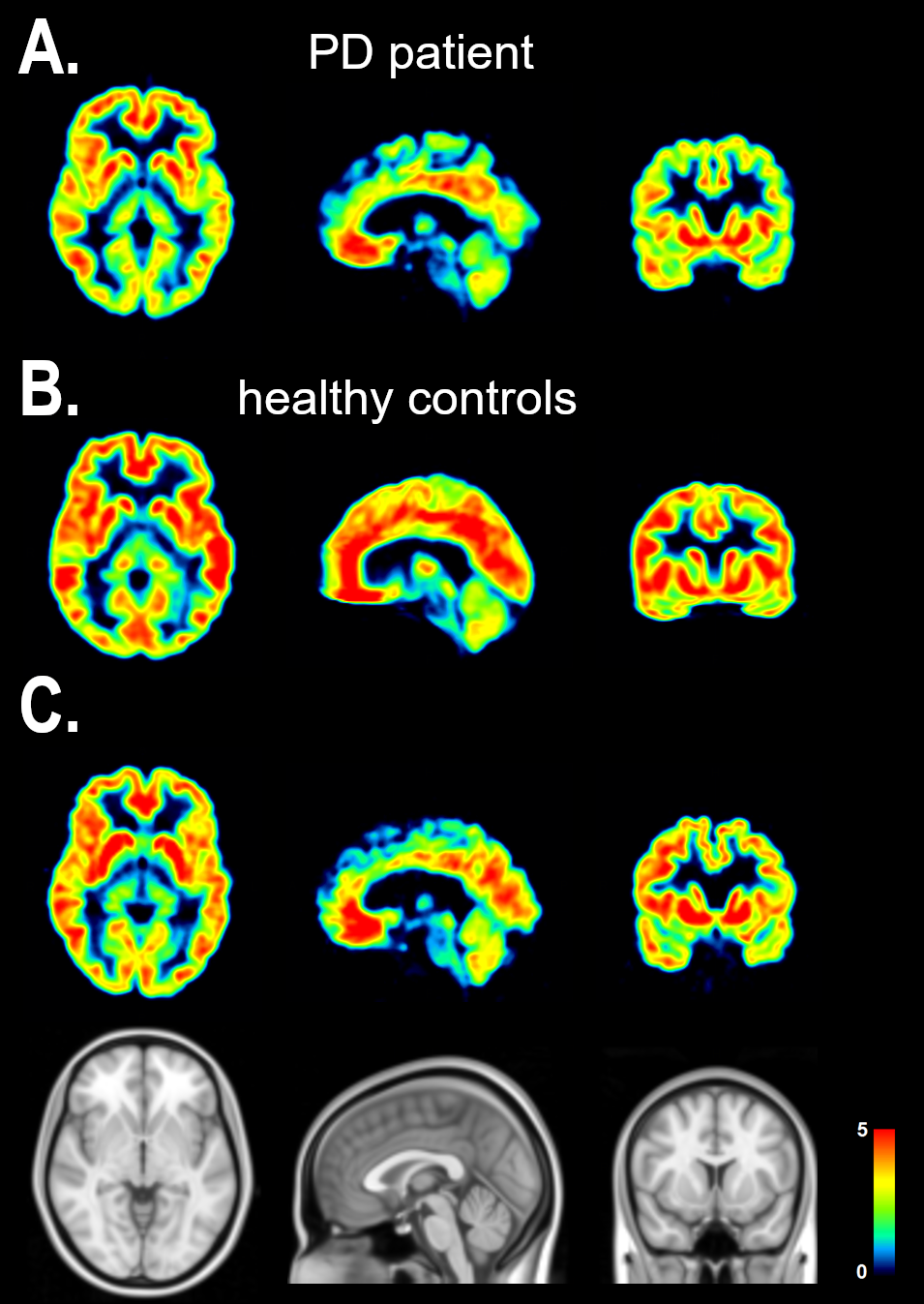
Method: In vivo imaging: Two healthy controls (HC1: female, 50y; HC2: male, 67y) and one PD patient (66y, 15y disease duration, MDS-UPDRS-III=34, H&Y=3) were scanned. PET scans were acquired on a GE Discovery MI PET-CT. The radiotracer was injected via bolus injection (range 183-191MBq), and emission data acquired for 120 min with arterial blood collection. Low dose CT images were used for attenuation correction, and dynamic image reconstruction was performed using filtered back-projection. PET images were matched to the subject’s T1-weighted MRI and processed using Pmod 4.2. For this analysis, we applied a Simplified Reference Tissue Model 2 (SRTM2) to compute BPND values with the Centrum Semiovale as reference region (5).
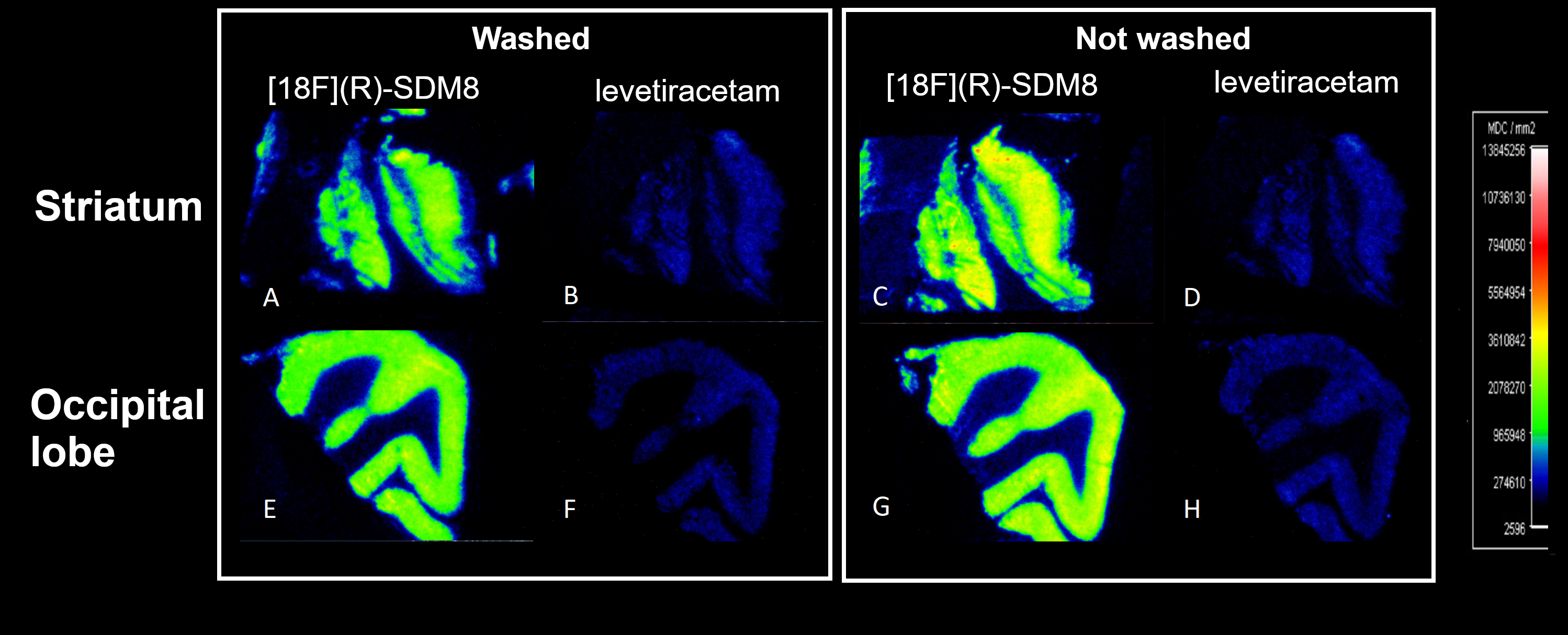
Postmortem imaging: The [18F](R)-SDM8 in vitro binding was assessed in the striatum and occipital lobe of a MSA patient. Sections were sliced at 10 µm and stored at -80°C. Slides were thawed and dried at room temperature for at least 30 minutes. Tissue was incubated for 1h with [18F](R)-SDM8 (2nM and 12,400 mCi/µmole) or with 200µM levetiracetam.
Results: Whole-brain BPND maps for the healthy controls and PD are shown in Fig.1 displaying distribution of BPND in the cortical and subcortical regions. Fig.2 shows the postmortem in vitro binding in sections of the striatum and occipital lobe of the MSA patient with [18F](R)-SDM8 and following levetiracetam.
Conclusion: The preliminary data with the [18F] labeled SV2A tracer displayed good in vivo binding distribution properties in PD, and postmortem MSA patient. The use of the [18F]SynVesT-1 tracer is a promising tool for synaptic density quantification in parkinsonism.
References: 1. Matuskey D, Tinaz S, Wilcox KC, Naganawa M, Toyonaga T, Dias M, et al. Synaptic Changes in Parkinson Disease Assessed with in vivo Imaging. Ann Neurol. 2020;87(3):329–38.
2. Delva A, Van Weehaeghe D, Koole M, Van Laere K, Vandenberghe W. Loss of Presynaptic Terminal Integrity in the Substantia Nigra in Early Parkinson’s Disease. Mov Disord. 2020;35(11):1977–86.
3. Wilson H, Pagano G, de Natale ER, Mansur A, Caminiti SP, Polychronis S, et al. Mitochondrial Complex 1, Sigma 1, and Synaptic Vesicle 2A in Early Drug-Naive Parkinson’s Disease. Mov Disord. 2020;35(8):1416–27.
4. Naganawa M, Li S, Nabulsi N, Henry S, Zheng MQ, Pracitto R, et al. First-in-Human Evaluation of 18 F-SynVesT-1, a Radioligand for PET Imaging of Synaptic Vesicle Glycoprotein 2A. J Nucl Med. 2021;62(4):561–7.
5. Naganawa M, Gallezot JD, Li S, Nabulsi NB, Henry S, Zheng MQ, et al. Noninvasive quantification of 18F-SynVesT-1 binding using simplified reference tissue model 2. [Abstract] Society of Nuclear Medicine and Molecular Imaging (SNMMI). 2022.
To cite this abstract in AMA style:
C. Uribe, KL. Desmond, R. Raymond, K. Smart, A. Reilhac, A. Mena, AE. Lang, G. Kovacs, N. Vasdev, AP. Strafella. In vivo synaptic density in parkinsonism [abstract]. Mov Disord. 2022; 37 (suppl 2). https://www.mdsabstracts.org/abstract/in-vivo-synaptic-density-in-parkinsonism/. Accessed December 18, 2025.« Back to 2022 International Congress
MDS Abstracts - https://www.mdsabstracts.org/abstract/in-vivo-synaptic-density-in-parkinsonism/


