Category: Neuroimaging (Non-PD)
Objective: To assess the relationship between synaptic density and functional connectivity in the primary tauopathy of Progressive Supranuclear Palsy (PSP).
Background: PSP is a debilitating neurocognitive disorder, associated with 4R tau aggregation. Synaptic loss and abnormal functional connectivity (FC) are proposed to underlie cognitive symptoms, even preceding atrophy. However, the relationship between synaptic density and FC in PSP is unknown. Here, we assess the relationship between in vivo synaptic density using [11C]UCB-J PET, and FC derived from resting state function MRI (rsfMRI) graph metrics.
Method: 36 participants were recruited: 18 with probable PSP-Richardson’s syndrome, and 18 age-/sex-/education-matched controls. Dynamic PET imaging quantified regional [11C]UCB-J non-displaceable binding potentials (BPND). Graph metrics (weighted degree: an index of the strength and number of local connections) were extracted from each nodal brain region in functional networks, constructed from rs-fMRI data. PET and regional connectivity data were normalised against controls, averaged across patients and entered a linear regression model with age as a covariate.
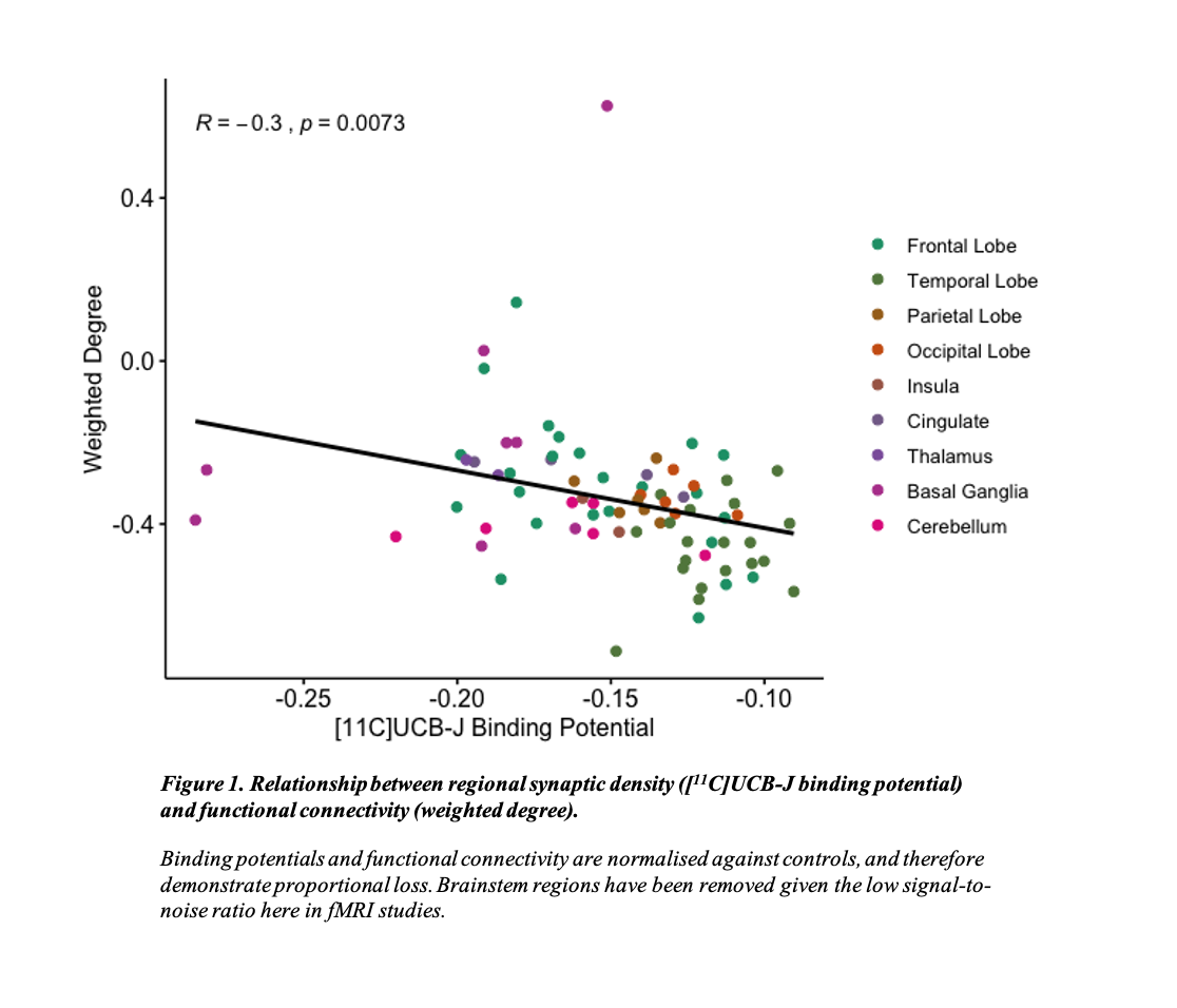
Results: Compared to controls, patients with PSP had significant widespread synaptic loss as measured by [11C]UCB-J PET in both cortical and subcortical regions [1], and abnormal weighted degree. Across all brain regions, stronger local connectivity related to more severe synaptic loss (beta= 3.6, t= -3.8, p = 0.0003) (Figure 1).
Conclusion: Our results suggest an in vivo link between synaptic loss and FC, in a disease where the primary burden of neuropathology and atrophy is sub-cortical. We firstly suggest that the extent of connectivity loss is dependent on baseline synaptic density, such that, for example in the frontotemporal cortex with a higher baseline synaptic density, connectivity loss is greatest, whereas in subcortical areas with lower baseline synaptic density, connectivity is relatively preserved. Second, we propose that our observation might represent diaschisis, with a loss of trophic support to synapses from long-range cortico-cortical connectivity, only partially compensated by an increase in short-range subcortical connectivity. Future longitudinal imaging will inform the directionality of mediation of these effects and their relationship to functional decline.
References: [1] Holland N, Jones PS, Savulich G, et al. Synaptic Loss in Primary Tauopathies Revealed by [11C]UCB-J Positron Emission Tomography. Mov Disord. 2020;35(10):1834-1842. doi:10.1002/mds.28188
To cite this abstract in AMA style:
N. Holland, T. Rittman, T. Cope, A. Perry, S. Jones, G. Savulich, M. Malpetti, T. Fryer, F. Aigbirhio, J. O'Brien, J. Rowe. In vivo relationship between synaptic density and functional connectivity in progressive supranuclear palsy: a multimodal [11C]UCB-J PET and resting state fMRI study. [abstract]. Mov Disord. 2021; 36 (suppl 1). https://www.mdsabstracts.org/abstract/in-vivo-relationship-between-synaptic-density-and-functional-connectivity-in-progressive-supranuclear-palsy-a-multimodal-11cucb-j-pet-and-resting-state-fmri-study/. Accessed April 22, 2025.« Back to MDS Virtual Congress 2021
MDS Abstracts - https://www.mdsabstracts.org/abstract/in-vivo-relationship-between-synaptic-density-and-functional-connectivity-in-progressive-supranuclear-palsy-a-multimodal-11cucb-j-pet-and-resting-state-fmri-study/

