Session Information
Date: Thursday, June 8, 2017
Session Title: Parkinson's Disease: Neuroimaging And Neurophysiology
Session Time: 1:15pm-2:45pm
Location: Exhibit Hall C
Objective: The objective of the present work was to evaluate in vivo with high resolution positron emission tomography (PET), the relative loss of the dopamine transporter (DAT) in the axonal terminals as compared with the cell bodies and axons, in early stages of Parkinson´s disease (PD).
Background: DAT is enriched in nigrostriatal dopaminergic neuron and it regulates the dopamine-related signaling. To date the nigrostriatal terminals in the striatum have been the main target for DAT imaging in early stages of PD (1). DAT is enriched at the level of the striatal terminals and on cell bodies of dopaminergic neurons in the substantia nigra but also in dendritic and axonal plasma membranes (2).
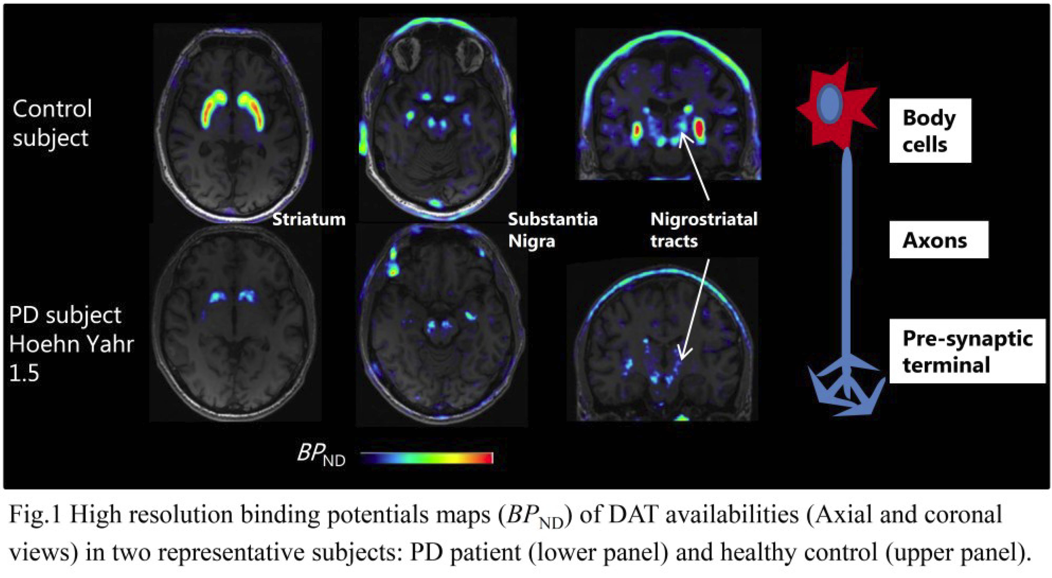
Methods: Nineteen PD patients (14M/5F, 61.8±8.4y, disease duration 2.9±2.6y; UPDRS-m: 19.4±6.5) and 14 control subjects (CS) (12M/2F, 60.6±6.8y) underwent PET measurements with the DAT radioligand 18F-FE-PE2I using the high resolution research tomography (HRRT) system. Voxel-based BPND maps were generated for all subjects (fig.1). Template-based regions of interest (ROIs) based on DAT binding pattern and anatomical information from MRI were first delineated for the substantia nigra (SN), the striatum (STR) and for the nigro-striatal (NST), nigro-pallidal (NPT) and nigro-thalamic (NTT) tracts. The ROIs were then applied to each parametric image to obtain individual BPND estimates. Differences between groups were assessed with unpaired t-test (p<0.05) and by calculating the effect size (Cohen’s d).
Results: In PD patients, significantly lower BPND values compared with CS (p<0.001) were observed in STR (1.7±0.7 vs 4.0±0.5) and SN (0.6±0.1 vs 0.8±0.1). No statistically significant differences of DAT availability between groups were measured along the tracts (PD: 0.41±0.1, CS: 0.44±0.1). Cohen´s d between groups (PD vs HC) were larger for the STR (3.2), moderate for the SN (1.8) and small for the tracts (0.2).
Conclusions: The findings of this study demonstrate that in early PD DAT availability is mainly decreased in the striatal terminals and to a much lower degree at the level of the cell bodies in the SN and preserved along the tracts. These results confirm earlier reports from post-mortem studies suggesting that in early PD a large proportion of dopaminergic neurons in the SN are still viable.
References: 1. Marek K, Innis R, van Dyck C, Fussell B, Early M, Eberly S, et al. 2001. [123I]beta-CIT SPECT imaging assessment of the rate of Parkinson’s disease progression. Neurology. 57:2089–94.
2. Hersch SM, Hong YI, Heilman CJ, Edwards RH, Levey AI. 1997. Subcellular localization and molecular topology of the dopamine transporter in the striatum and substantia nigra. J Comp Neurol. 388:211–227.
To cite this abstract in AMA style:
P. Fazio, P. Svenningsson, Z. Cselenyi, C. Halldin, L. Farde, A. Varrone. In vivo mapping of nigro-striatal dopamine transporter (DAT) availability in early Parkinson´s disease patients using [18F]FE-PE2I and high-resolution positron emission tomography. [abstract]. Mov Disord. 2017; 32 (suppl 2). https://www.mdsabstracts.org/abstract/in-vivo-mapping-of-nigro-striatal-dopamine-transporter-dat-availability-in-early-parkinsons-disease-patients-using-18ffe-pe2i-and-high-resolution-positron-emission-tomography/. Accessed December 31, 2025.« Back to 2017 International Congress
MDS Abstracts - https://www.mdsabstracts.org/abstract/in-vivo-mapping-of-nigro-striatal-dopamine-transporter-dat-availability-in-early-parkinsons-disease-patients-using-18ffe-pe2i-and-high-resolution-positron-emission-tomography/

