Category: Parkinson's Disease: Neuroimaging
Objective: To assess two aspects of the noradrenergic system using multimodal in vivo imaging in a sample of Parkinson’s disease (PD) patients and healthy controls (HC): the pigmented cell bodies of the locus coeruleus (LC) with neuromelanin sensitive MRI and the α2-adrenergic receptors (ARs) density with [11C]yohimbine PET.
Background: Degeneration of the noradrenergic system is nowadays considered as a pathological hallmark of PD [1]. In addition, both antiparkinsonian and neuroprotective properties of the noradrenergic system have been suggested [2]. Yet, the therapeutic strategies targeting this system in PD are still currently limited as knowledge in the noradrenergic system in vivo are scarce.
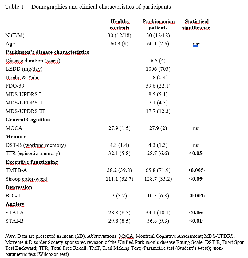
Method: 30 PD patients and 30 HC age- and gender-matched subjects were included. [Table 1]
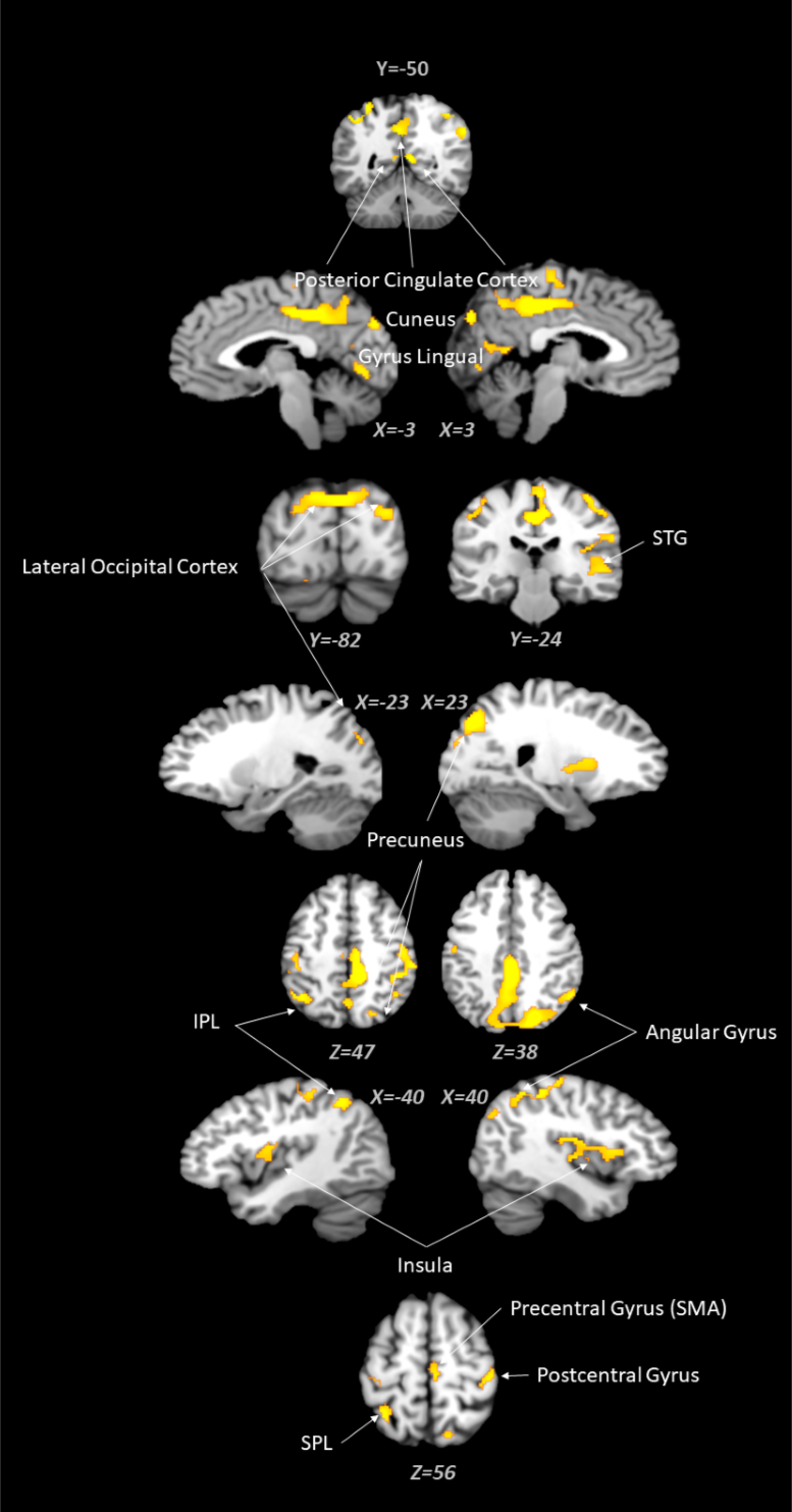
Results: A reduction in [11C]yohimbine binding in PD patients compared to HC was observed in widespread cortical regions encompassing the parietal lobe, the posterior cingulate cortex (PCC), the temporal and occipital cortex as well as the motor cortices [Figure 1]. In parallel, PD patients showed a reduced signal intensity in LC compared with HC (p<0.001). Interestingly, worse performances in executive functioning within the PD group were correlated with a decrease in LC signal intensity (e.g. TMTB-A: r=-0.46), and an increase in [11C]yohimbine binding within specific areas including the right pre- and post-central gyrus and occipital cortex (r=0.38). We also showed that both higher anxiety and depressive symptoms were associated with reduced [11C]yohimbine binding in the left PCC (r=-0.32 and -0.51 respectively) and the right inferior frontal gyrus (r=-0.37 and -0.38 respectively). Finally, the loss of pigmented neurons in the LC was negatively correlated with motor impairments (e.g. UPDRS-III: r=-0.48).
Conclusion: Our results support the existence of substantial PD-related declines in the integrity of the noradrenergic system and highlight its role in the PD pathophysiology. In particular, the decline of LC signal intensity was associated with both executive and motor impairments while the decrease in [11C]yohimbine binding was mainly associated with the magnitude of depressive, anxious and executive symptoms.
References: [1] Laurencin C, Lancelot S, Gobert F, Redouté J, Mérida I, Iecker T, et al. Modeling [11C]yohimbine PET human brain kinetics with test-retest reliability, competition sensitivity studies and search for a suitable reference region. Neuroimage. 2021 Oct 15;240:118328.
[2] Paredes-Rodriguez E, Vegas-Suarez S, Morera-Herreras T, De Deurwaerdere P, Miguelez C. The Noradrenergic System in Parkinson’s Disease. Front Pharmacol. 2020;11:435.
[3] O’Neill E, Harkin A. Targeting the noradrenergic system for anti-inflammatory and neuroprotective effects: implications for Parkinson’s disease. Neural Regeneration Research. 2018;13(8):1332.
To cite this abstract in AMA style:
C. Laurencin, S. Lancelot, M. Huddlestone, S. Brosse, I. Merida, N. Costes, J. Redouté, S. Thobois, P. Boulinguez, B. Ballanger. Imaging the noradrenergic system in Parkinson’s disease: A [11C]Yohimbine PET and neuromelanin MRI study [abstract]. Mov Disord. 2023; 38 (suppl 1). https://www.mdsabstracts.org/abstract/imaging-the-noradrenergic-system-in-parkinsons-disease-a-11cyohimbine-pet-and-neuromelanin-mri-study/. Accessed December 16, 2025.« Back to 2023 International Congress
MDS Abstracts - https://www.mdsabstracts.org/abstract/imaging-the-noradrenergic-system-in-parkinsons-disease-a-11cyohimbine-pet-and-neuromelanin-mri-study/


