Category: Surgical Therapy: Parkinson's Disease
Objective: To evaluate the utility of image-guided DBS programming; to determine the correlation between optimal DBS electrode placement predicted by imaging-guided software and monopolar review in Parkinson’s disease (PD) patients with DBS.
Background: The current paradigm for deep brain stimulation (DBS) programming, monopolar review, is a time-consuming process – from about 40 minutes[1] up to 140 minutes[2] for bilateral leads. Initial programming is performed in an off-medication state, causing patient discomfort.
New imaging software allows visualization of each electrode contact in a patient-specific anatomical model based on CTs and MRIs. Image-guided programming (IGP) for DBS in PD patients achieved similar motor symptom control after 4 weeks and can significantly reduce programming time[1,2].
Method: A software algorithm (Brainlab) fused preoperative MRIs with postoperative CTs to create a patient-specific anatomical atlas and visualize each lead in STN or GPI. Two reviewers independently assessed these fused images for 70 patients who had DBS surgery for PD. Using these images, each contact level was stratified into one of four categories based on percent submersion in the target structure: full (100%), majority (50-99%), partial (1-50%), no (0%) submersion. Retrospective chart review determined the correlation between IGP and monopolar review.
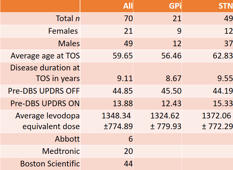
Results: Analysis included 70 patients, 21 GPi and 49 STN. Total number of leads was 139: 41 GPi and 98 STN.
Baseline demographics in Table 1. Males represented a higher proportion. STN patients were older at the time of surgery.
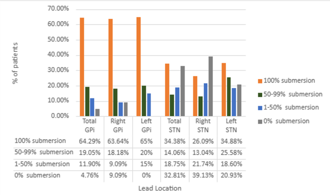
Results in Table 2. The majority of GPi contact levels (64%) deemed best during initial monopolar review correlated with those predicted by the software and had full submersion in GPi. For left STN leads, 35% of contact levels used in monopolar review had full submersion; the right leads had a largest percentage (39%) with no submersion.
Conclusion: IGP correlated well with monopolar review for the majority of GPi leads, but less robustly for STN leads. Potential explanations include: difference in target size; sequence effect – the correlation was lowest for right STN leads (typically implanted second in our institution); intraoperative brain shift. It is also possible that imaging software cannot yet account for lead migration and electrode impedances. Patient-specific anatomical programming can be a useful tool, which must be balanced with clinical response.
References: 1. Pourfar MH, Mogilner AY, Farris S, Giroux M, Gillego M, Zhao Y, Blum D, Bokil H, Pierre MC. Model-Based Deep Brain Stimulation Programming for Parkinson’s Disease: The GUIDE Pilot Study. Stereotact Funct Neurosurg 2015;93(4):231-9.
2. Pavese N, Tai YF, Yousif N, Nandi D, Bain PG. Traditional Trial and Error versus Neuroanatomic 3-Dimensional Image Software-Assisted Deep Brain Stimulation Programming in Patients with Parkinson Disease. World Neurosurg. 2020 Feb;134:e98-e102.
To cite this abstract in AMA style:
S. Marmol, J. Moll, H. Bullock, I. Haq, J. Jagid, C. Luca. Image-guided programing in useful in DBS for Parkinson’s disease [abstract]. Mov Disord. 2023; 38 (suppl 1). https://www.mdsabstracts.org/abstract/image-guided-programing-in-useful-in-dbs-for-parkinsons-disease/. Accessed October 15, 2025.« Back to 2023 International Congress
MDS Abstracts - https://www.mdsabstracts.org/abstract/image-guided-programing-in-useful-in-dbs-for-parkinsons-disease/


