Session Information
Date: Wednesday, June 7, 2017
Session Title: Myoclonus
Session Time: 1:15pm-2:45pm
Location: Exhibit Hall C
Objective: We report the findings of a patient presenting with palatal myoclonus (PM) several months after suffering from an Intracranial Hemorrhage (ICH) in order to demonstrate the unique neuroanatomical location of PM.
Background: PM is characterized by a 1-3 Hz frequency oscillation of the palate. It can be appreciated with vocalization as well as rest. PM has been demonstrated to result from a lesion within the dentate-rubro-olivary pathway (Guillain-Mollaret triangle) due to, most commonly, a cerebrovascular or demyelinating lesion. There may be subsequent associated development of hypertrophic olivary degeneration (HOD) observed on MRI. Further motor symptoms may be appreciated depending on location of this lesion within the pathway.
Methods: 72 year old female professional opera singer presented to our clinic with complaints inability to sing, left arm tremor with associated stiffness, and slowness of movements over the past six months. Eleven months prior to the onset of these symptoms, she had a left cerebellar ICH with residual vertigo and gait ataxia. Neurological exam revealed a vibratory quality to her voice with inability to sustain vocal notes and left hand wing-beating tremor present at rest, posture, and action. Additionally, she had bilateral, left worse than right bradykinesia and rigidity. She was started on carbidopa-levodopa 25/100 mg TID with moderate improvement in her parkinsonism, however the PM did not improve.
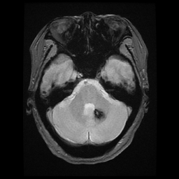
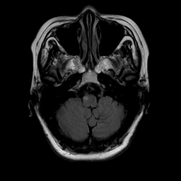
Results: Blood count, blood chemistry including liver enzymes, and thyroid stimulating hormone were normal. LDL cholesterol level was elevated at 145 mg/dl. MRI Brain revealed a focal area of high T2 signal intensity and enlargement of the right medullary olive and an area of susceptibility on GRE of the contralateral dentate nucleus from the ICH [Figure 1]. The left ICH and enlargement of the right medullary olive suggesting hypertrophic olivary degeneration with disruption of dentato-rubro-olivary pathway [Figure 2] was thought to cause the presentation of PM, Holmes (or cerebellar) tremor, and secondary parkinsonism.
Conclusions: Disruption of the dentate-rubro-olivary classically presents with palatal myoclonus with associated features with imaging demonstrating a lesion within the pathway and secondary hypertrophy of the inferior olive. These symptoms may present from months to years after the initial insult emphasizing the importance of awareness of this delayed presentation.
References: Vidailhet, M., Jedynak, C., Pollak, P., Agid, Y. (1998). Pathology of Symptomatic Tremors. Movement Disorders Journal, 13(3), 49-54. http://dx.doi.org/10.1002/mds.870131309
Deuschl, G., Toro, C., Valls-Sole, J., Zeffiro, T., Zee, D., Hallett, M. (1994). Symptomatic and essential palatal tremor: Clinical, physiological and MRI analysis. Brain, 117 (4), 775-788 http://dx.doi.org/10.1093/brain/117.4.775
To cite this abstract in AMA style:
G. Moreno, K. Ng, A. Duffy. Hypertrophic Olivary Degeneration [abstract]. Mov Disord. 2017; 32 (suppl 2). https://www.mdsabstracts.org/abstract/hypertrophic-olivary-degeneration/. Accessed April 20, 2025.« Back to 2017 International Congress
MDS Abstracts - https://www.mdsabstracts.org/abstract/hypertrophic-olivary-degeneration/


