Category: Rare Genetic and Metabolic Diseases
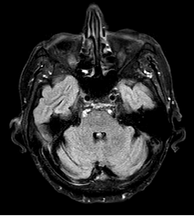
Objective: We present a case of Marchiafava-Bignami Disease (MBD), where reversible hyperintensity in Middle Cerebellar Peduncle (MCP-sign) is observed.
Background: MCP sign is an infrequent finding in brain MRI, usually in the context of neurodegenerative pathologies, is considered a classic sign (and major diagnostic criteria) of fragile X-associated tremor ataxia syndrome. Its description in other entities is anecdotal.
Method: A 59-year-old male, with 30 years history of alcoholism, presents subacute gait disturbance and cognitive deterioration, with progressive worsening over 6 months, requiring hospital admission.
On examination, we observe somnolence, dysarthria, cerebellar and sensory ataxia, and cognitive impairment (recovery-memory deficit, alteration of processing speed and executive-attentional functions). No tremor or other movement disorders were observed during examination.
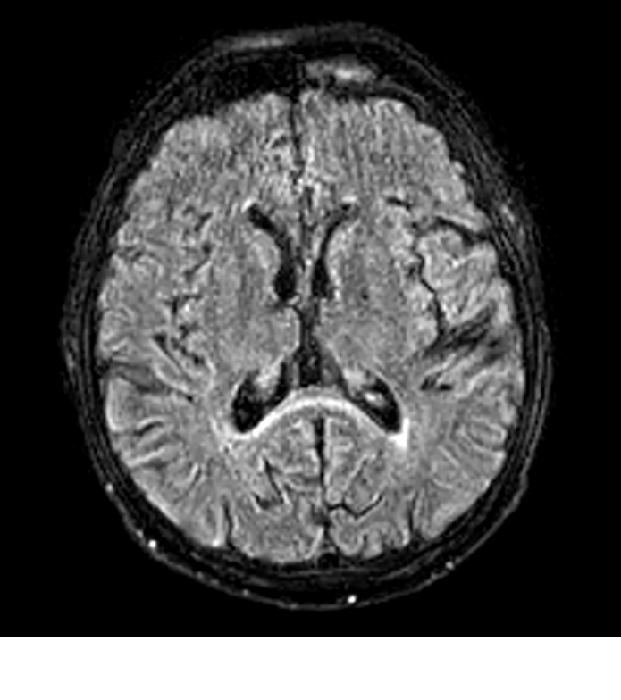
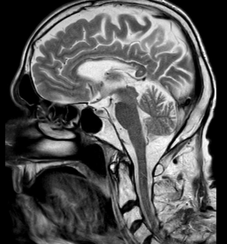
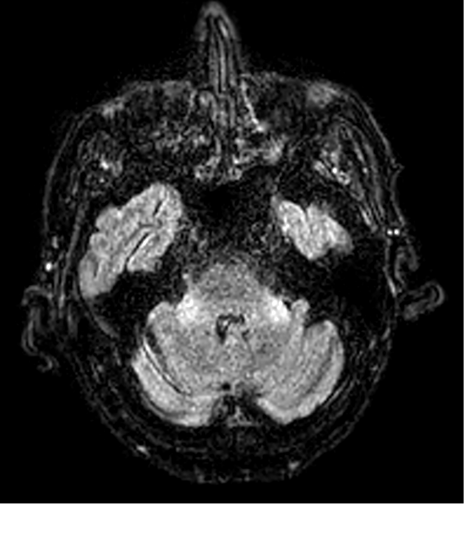
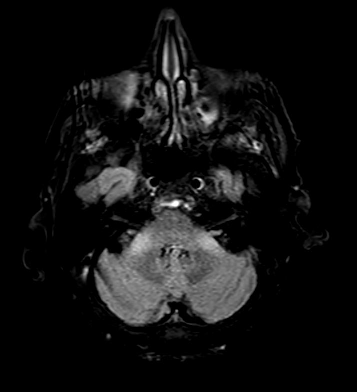
Brain MRI showed global thinning of the corpus callosum, T2 hyperintensity at the splenium (Figure-1,2) and MCP-sign (Figure-3,4); findings described in MBD.
A comprehensive analytical study showed deficiency of folic acid and thiamine. Genetic study of the FMR1 premutation showed no alterations.
Results: Final diagnostic judgement was probable MBD type B, based on clinical and radiological findings. Treatment with vitamin supplementation showed progressive clinical improvement (being able to walk with a walker). A follow-up brain MRI, 2 months after hospital discharge, highlights the resolution of MCP sign (Figure-5), but persistence of the findings in the corpus callosum.
Conclusion: MBD is an infrequent pathology (probably underdiagnosed), associated with alcoholism and malnutrition, characterized by demyelinating lesions involving the corpus callosum. The few cases described demonstrate a highly variable clinical presentation. Today, the diagnosis is based on neuroimaging findings: hyperintense lesions in corpus callosum, hemispheric white matter, etc. The MCP-sign has been described only in a few case-reports. In our case, good clinical and radiological response (with resolution of the MCP-sign) stands out after vitamin supplementation.
The presence of MCP-sign should also suggest the possibility of MBD, and early vitamin and nutritional assessment should be performed, being an infrequent but treatable entity, with a potentially fatal course in the absence of treatment.
References: – S. Bellido, MD, I. Navas, MD, M.A. Aranda, MD, R. Ginestal, MD, B. Venegas, MD. Unusual MRI findings in a case of Marchiafava Bignami disease. Neurology 2012; 78; 1537 – C.-S. Tung. S.-L. Wu et al. Marchiafava-Bignami Disease with Widespread Lesions and Complete Recovery. American Journal of Neuroradiology September 2010, 31 (8) 1506-1507
To cite this abstract in AMA style:
D. López Domínguez, M. Puig Casadevall, G. álvarez Bravo. Hyperintensity in Middle Cerebellar Peduncles, an infrequent and reversible finding in Marchiafava-Bignami Disease. [abstract]. Mov Disord. 2021; 36 (suppl 1). https://www.mdsabstracts.org/abstract/hyperintensity-in-middle-cerebellar-peduncles-an-infrequent-and-reversible-finding-in-marchiafava-bignami-disease/. Accessed December 24, 2025.« Back to MDS Virtual Congress 2021
MDS Abstracts - https://www.mdsabstracts.org/abstract/hyperintensity-in-middle-cerebellar-peduncles-an-infrequent-and-reversible-finding-in-marchiafava-bignami-disease/

