Session Information
Date: Wednesday, September 25, 2019
Session Title: Neuroimaging
Session Time: 1:15pm-2:45pm
Location: Les Muses Terrace, Level 3
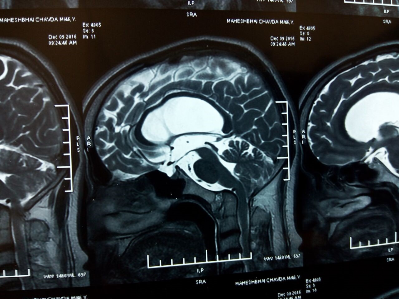
Objective: To report a case of Craniovertebral junction anomaly leading to a “Humming bird” sign on magnetic resonance imaging [MRI].
Background: Humming bird sign is used to describe sagittal view magnetic resonance image (MRI) of brainstem, looking like a bird. It occurs due to midbrain atrophy without pontine atrophy. It has been described to have high sensitivity and specificity for progressive supranuclear palsy (PSP) but here we present a case of Craniovertebral junction anomaly with Hummingbird sign on MRI leading to misdiagnosis.
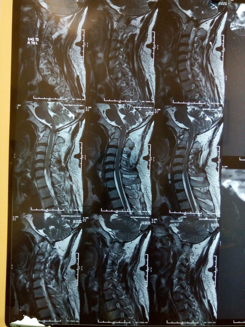
Method: A 49 year male presented with insidious onset gradually progressive imbalance, with slowness and stiffness of all limbs for 3-4 years. He had mild rigidity, with restricted vertical eye movements with generalized hyperreflexia. He was initially evaluated at other centre and diagnosed as PSP based on humming bird sign on MRI brain.[fig 1] However, his detailed examination revealed short neck, low hairline, predominant spasticity rather rigidity. He actually had chronic nuclear left third nerve palsy leading to bilateral ptosis with left esotropia. A detailed review of MRI demonstrated occipitalisation of C1 vertebra, congenital fusion of C2 vertebra, odontoid process hypoplasia, Atlanto-Axial Subluxation causing holoprosencephaly, moderate dilatation of 3rd Ventricle and Lateral Ventricles [fig 2]. This chronic compression of midbrain due to dialted 3rd ventricle was causing midbrain atrophy with preserved pons forming the Humming bird sign on sagittal MRI.
Results: Hummingbird is commonly considered to be associated with PSP, although it has also been described with Fragile X Associated Tremor/Ataxia syndrome and normal pressure hydrocephalus. We report a Humming Bird sign in cranio-vertebral junction anomaly. The postulated mechanism for this phenomenon is Atlanto-Axial Subluxation causing holoprosencephaly, moderate dilatation of 3rd Ventricle and Lateral Ventricles. This chronic compression of midbrain due to dialted 3rd ventricle was causing midbrain atrophy with preserved pons forming the Humming bird sign on sagittal MRI.
Conclusion: A detailed clinical examination and detailed review of imaging findings is essential. Just considering a radiological finding (Hummingbird sign) without detailed examination can lead to misdiagnosis of PSP as demonstrated in our case.
To cite this abstract in AMA style:
D. Desai, D. Pandya, S. Desai. Humming bird sign in craniovertebral junction anomaly leading to misdiagnosis of progressive supranuclear palsy [abstract]. Mov Disord. 2019; 34 (suppl 2). https://www.mdsabstracts.org/abstract/humming-bird-sign-in-craniovertebral-junction-anomaly-leading-to-misdiagnosis-of-progressive-supranuclear-palsy/. Accessed January 1, 2026.« Back to 2019 International Congress
MDS Abstracts - https://www.mdsabstracts.org/abstract/humming-bird-sign-in-craniovertebral-junction-anomaly-leading-to-misdiagnosis-of-progressive-supranuclear-palsy/


