Session Information
Date: Thursday, June 8, 2017
Session Title: Parkinson's Disease: Neuroimaging And Neurophysiology
Session Time: 1:15pm-2:45pm
Location: Exhibit Hall C
Objective: To identify patterns of grey matter (GM) abnormalities in patients with Parkinson’s disease (PD) with different cognitive profiles.
Background: Cognitive symptoms occur already in early stages of PD.
Methods: Patient subgroups (total N=124) were previously defined by a data-driven approach, resulting in four cognitive profiles: cognitively intact patients, patients with slight mental slowing only, patients with a dysexecutive syndrome and patients with severe cognitive dysfunction in all cognitive domains. We used optimized voxel-based morphometry to compare GM volume in all groups to the cognitively intact group and vertex-based morphometry to localize GM differences in cortical thickness (CT) and cortical folding (CF).
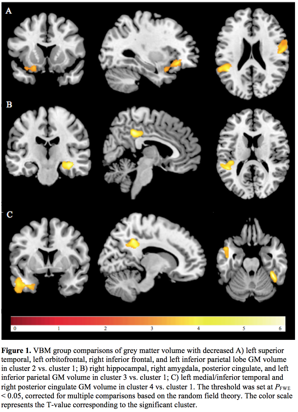
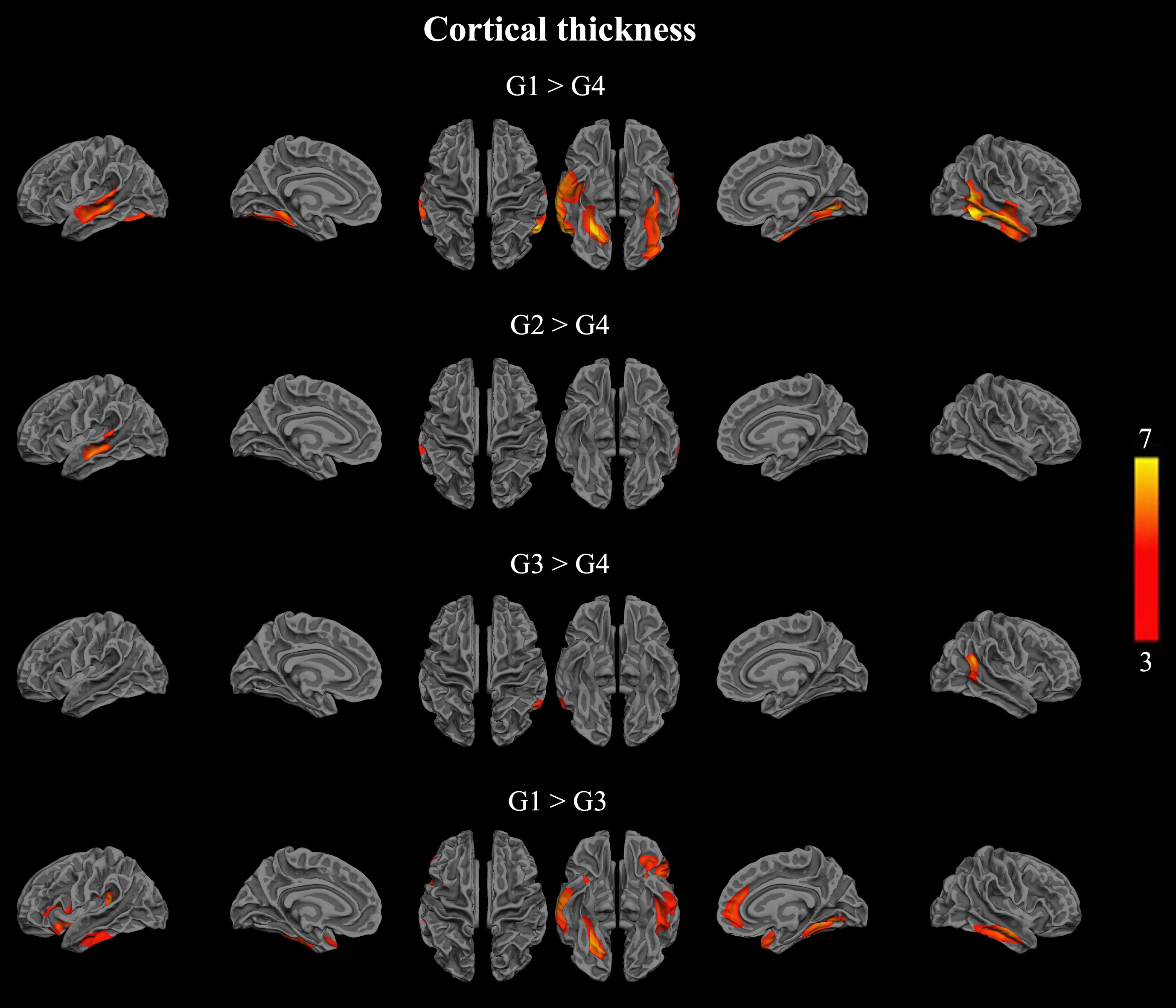
Results: Patients with slight mental slowing only showed reduced GM volume in temporal, parietal and frontal regions compared to cognitively intact patients, whereas patients with dysexecutive deficits showed reduced GM volume in the parietal-temporal lobe, posterior cingulate, bilateral hippocampus and amygdala. Additionally, these patients showed reduced CT in temporal, insular and anterior cingulate regions. Patients with severe cognitive deficits showed reduced GM volume in the left medial temporal lobe and left parahippocampal gyrus in addition to those regions affected in the dysexecutive group [figure1]. Furthermore, these patients showed reduced CT and CF in bilateral temporal regions and reduced CF in right precentral and inferior parietal gyrus, left insula and left rectus gyrus [figure 2 and 3]. These results were robust for differences in center, gender, and education. When corrected for age, only patients with severe cognitive deficits showed abnormalities in GM volume, CT and CF.
Conclusions: Reduced GM volume in temporal, parietal and frontal regions appeared to be an early neuroanatomical marker of cognitive decline in PD patients with reduced mental speed only. Hence, slowed mental speed may be considered as a first, yet crucial, sign of cognitive deterioration in PD. Age at evaluation appeared to be an important predictor of differences in GM indices. However, in patients with severe cognitive deficits, we found GM abnormalities unrelated to age that may resemble effects of disease processes in addition to aging, such as more widespread Lewy body pathology potentially with comorbid Alzheimer’s disease pathology.
To cite this abstract in AMA style:
A. Moonen, R. Lopes, A. Leentjens, A. Duits, K. Dujardin. Grey matter abnormalities associated with early cognitive decline in Parkinson’s disease [abstract]. Mov Disord. 2017; 32 (suppl 2). https://www.mdsabstracts.org/abstract/grey-matter-abnormalities-associated-with-early-cognitive-decline-in-parkinsons-disease/. Accessed January 4, 2026.« Back to 2017 International Congress
MDS Abstracts - https://www.mdsabstracts.org/abstract/grey-matter-abnormalities-associated-with-early-cognitive-decline-in-parkinsons-disease/


