Category: Parkinson's Disease: Neuroimaging
Objective: This study aimed to investigate the association of changed gray matter volume(GMV)with Freezing of gait(FOG) in Parkinson’s disease(PD).
Background: FOG is a characteristic gait disorder in PD.However, the pathogenesis of PD with FOG has not been elucidated.Current neuroimaging studies of PD with FOG are often variable or even partially conflicting, which reflect the heterogeneity and complexity of the phenomena.This study was based on voxel-based morphometry (VBM) analysis and combined with clinical scales to investigate the change of GMV in PD with FOG patients and its significance.
Method: This study included 54 PD without FOG(PD-NFOG)patients and 50 PD with FOG(PD-FOG)patients and 33 subjects as healthy control.Clinical characteristics,motor and non-motor symptom assessments and VBM analysis of 3D T1 weighted MRI were recorded for each group.Univariate statistical analysis was performed between the groups, followed by binary logistic regression and receiver operating characteristics(ROC)curves to identify clinical and neuroimaging biomarkers that may help predict the risk of PD-FOG.
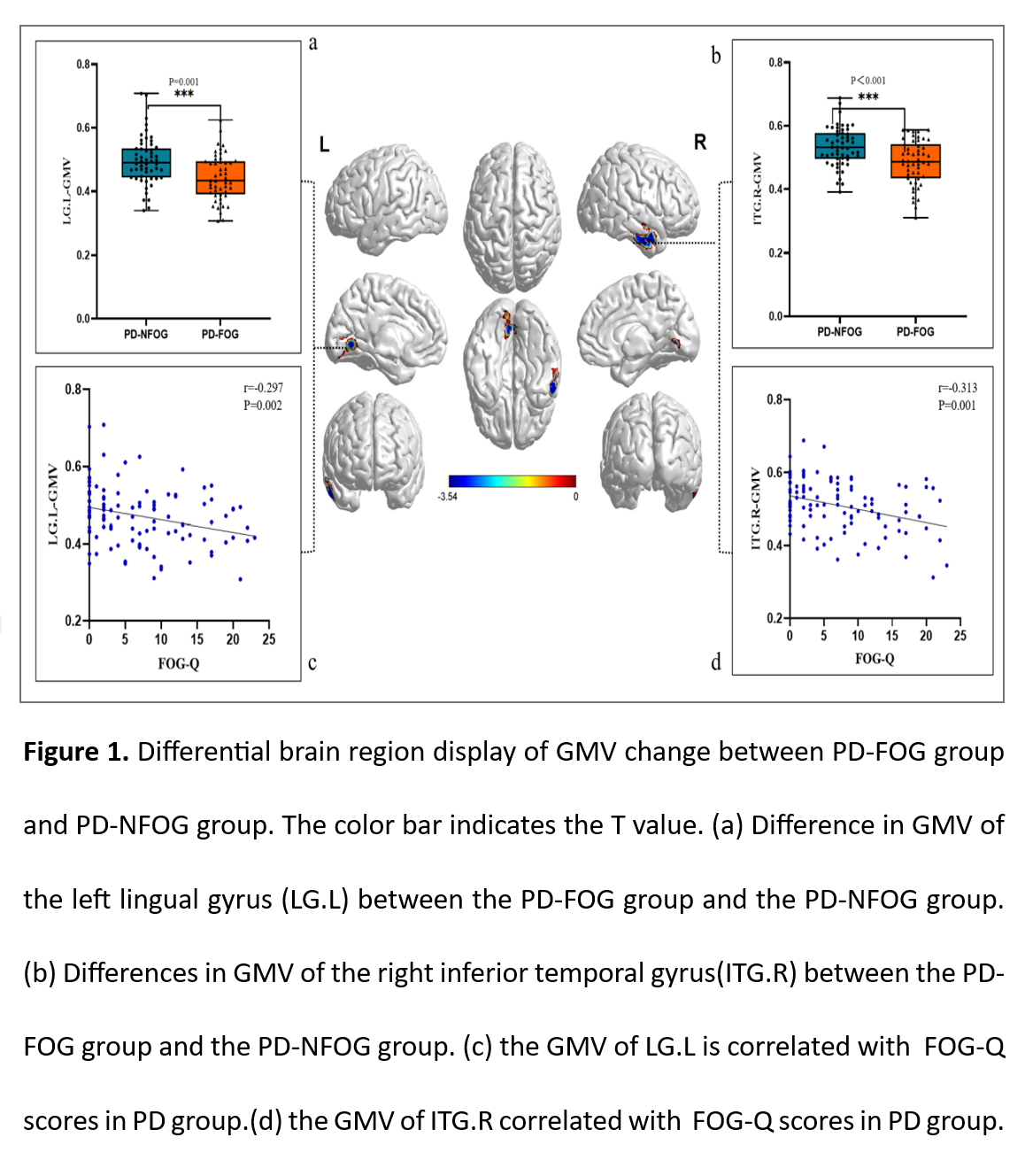
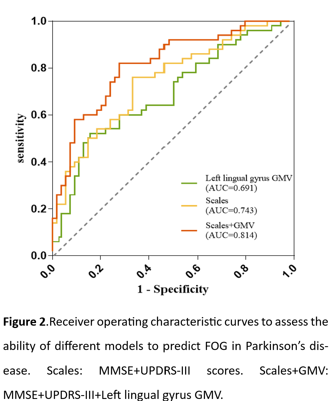
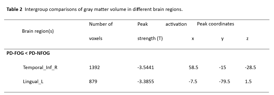
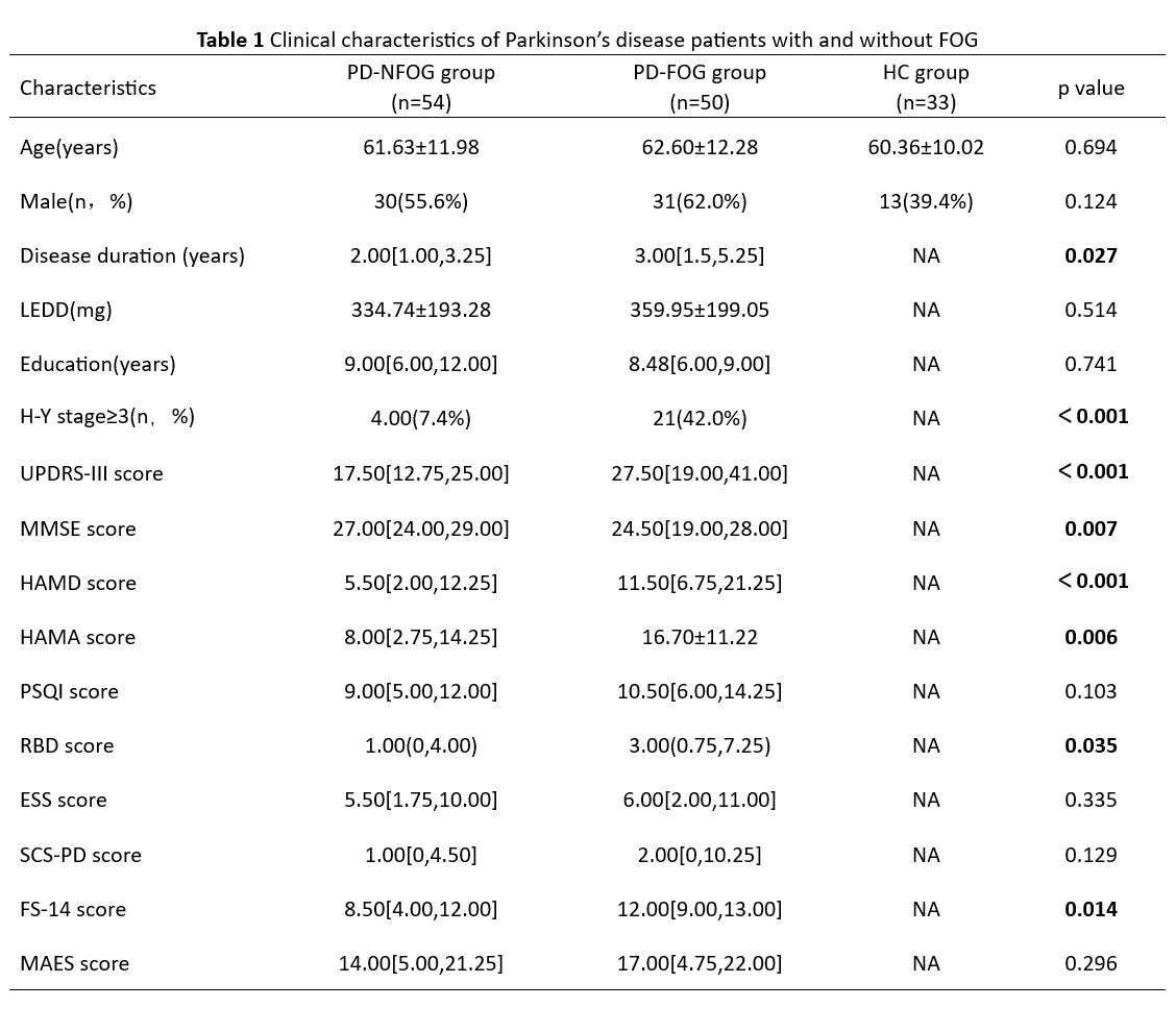
Results: The disease duration in the PD-FOG group was longer than the PD-NFOG group.The PD-FOG group in Unified Parkinson’s Disease Rating Scale part III(UPDRS-III),Hamilton Depression Scale(HAMD),Hamilton Anxiety Scale(HAMA),REM Sleep Behavior Disorder Screening Questionnaire(RBD-SQ),Fatigue Scale-14(FS-14),Freezing of gait-questionnaire(FOG-Q)scores and Hoehn-Yahr(H-Y)stage was higher than PD-NFOG group,and the Mini-mental State Examination(MMSE)scores were lower than PD-NFOG group[see table 1].Compared with PD-NFOG group,it was found that the GMV decreased in the right inferior temporal gyrus and the left lingual gyrus of PD-FOG groups[see Table 2, Figure 1].The combination of the GMV of left lingual gyrus and scales(MMSE+UPSRS-III)showed promising potential for predicting PD-FOG based on the area under the ROC curve (0.814,95% CI 0.733-0.896, p <0.001)[see Figure 2].
Conclusion: In conclusion,our study revealed differences in clinical features and GMV in specific brain regions in PD-FOG,suggesting that changes in GMV of the occipital and temporal regions associated with visual information processing may contribute to FOG.However,based on the differences in the results of current imaging studies,the comprehensive analysis of clinical features may improve the accuracy of PD-FOG risk prediction.
To cite this abstract in AMA style:
YX. Li, ZL. Luo, XL. Yang. Gray matter volume changes in occipital and temporal visual brain region are associated with freezing of gait in Parkinson’s [abstract]. Mov Disord. 2023; 38 (suppl 1). https://www.mdsabstracts.org/abstract/gray-matter-volume-changes-in-occipital-and-temporal-visual-brain-region-are-associated-with-freezing-of-gait-in-parkinsons/. Accessed December 15, 2025.« Back to 2023 International Congress
MDS Abstracts - https://www.mdsabstracts.org/abstract/gray-matter-volume-changes-in-occipital-and-temporal-visual-brain-region-are-associated-with-freezing-of-gait-in-parkinsons/


