Category: Parkinson's Disease: Neuroimaging
Objective: Our objective is to assess functional networks in PD mild cognitive impairment (PD-MCI) and their contribution to global connectivity combining Connectome ICA (connICA) and targeted resilience analysis.
Background: Brain network changes in PD-MCI are mostly unknown.
Method: 33 PD-MCI patients, 26 cognitively normal (PDCN), and 28 healthy controls (HC) underwent MRI. PD-MCI was diagnosed according to the MDS Task Force Guidelines (level II) [1]. T1-w and T2-w high resolution scans, and resting state BOLD fMRI images with standard monoband (TR=2s, 33 slices) and multiband (TR=800ms, 45 slices) GE-EPI images (3 mm isotropic voxels, matrix size= 64×64, TE= 28ms) were obtained. Brain parcellation was done using FreeSurfer based on Destrieux atlas (72 cortical and 8 subcortical regions/hemisphere). After preprocessing, 21 HC, 21 PDCN, and 23 PD-MCI subjects remained for analysis. FC matrices were computed based on Pearson’s correlation between 416 ROIs (Schaefer atlas plus subcortical areas of Destrieux atlas). A connICA was run in MELODIC with 65 independent FC-traits [2]. Using a linear mixed effect model and group-level Bayesian statistics [3], differences between groups were revealed. Network resilience was tested by eliminating nodes and recalculating global efficiency (GE). A partial least squares analysis [4] was done to assess the explained variance by the traits.
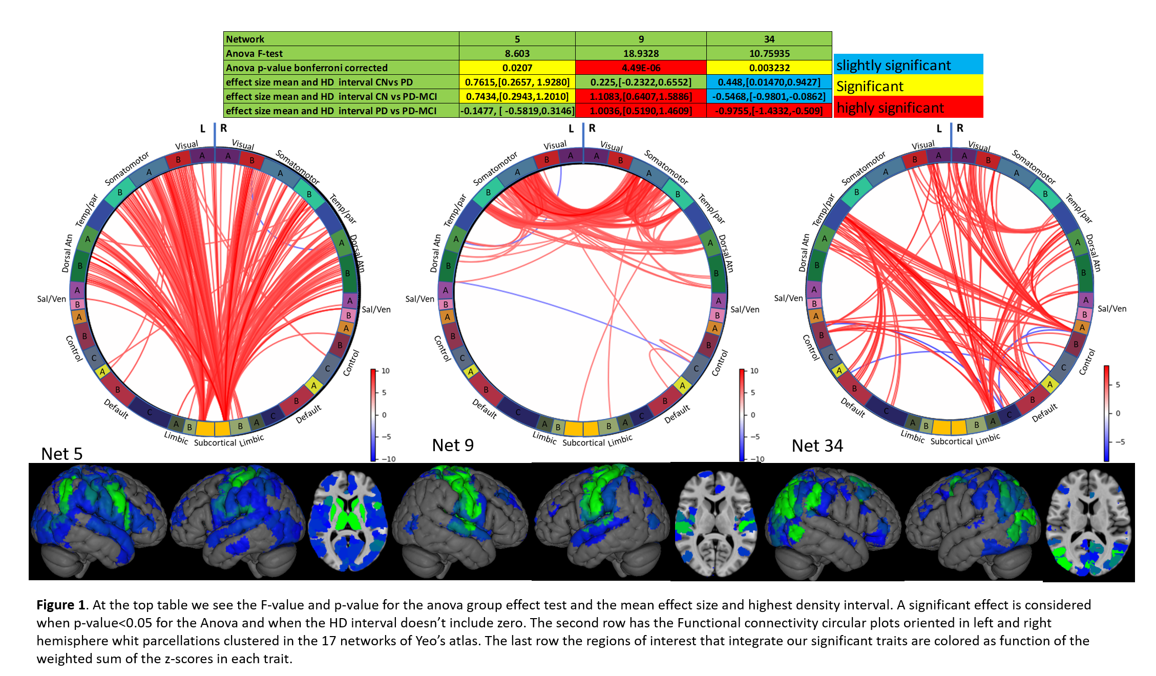
Results: Significant group differences were found for three traits (5, 9, and 34) (Table in Fig1). There was a significant effect size in FC-trait 5 in the comparisons HC vs. PDCN and HC vs. PD-MCI, and in FC-trait 9 for HC vs. PD-MCI and PDCN vs. PD-MCI. The targeted attack to these two networks (Fig2) showed a higher decrease of global efficiency (GE) in HC and PDCN while PD-MCI did not show such a decrease. This could reflect that connectivity of those networks is already deteriorated in PD-MCI. FC-trait 34, different across the 3 comparison groups, involves Angular gyrus and posterior cortical connections. The targeted analysis (Fig2) shows how deteriorating this trait negatively affects GE in PD-MCI. This could be explained by a key role of this functional network in cognitive reserve of PD-MCI patients.
Conclusion: Two Independent connectome traits were identified in the comparison PDCN vs PD-MCI. FC trait 34 mainly involving connections among posterior cortical areas might be of critical importance in cognitive reserve in PD-MCI patients.
References: [1] Litvan I, et al (2012). Diagnostic criteria for mild cognitive impairment in Parkinson’s disease: Movement Disorder Society Task Force guidelines. Movement Disorders vol. 27 pp. 349–56 [2] Amico E, et al (2018). The quest for identifiability in human functional connectomes. Scientific Report vol. 8 num 8254. [3] Kruschke JK (2013). Bayesian estimation supersedes the t test. Journal of Experimental Psychology vol. 142 pp. 573–603. [4] McIntosh AR, et al (2004). Partial least squares analysis of neuroimaging data: Applications and advances. In: NeuroImage supp1 pp. 250-263.
To cite this abstract in AMA style:
M. Delgado-Alvarado, V. Ferrer-Gallardo, I. Navalpotro-Gómez, S. Moia, M. Carreiras, C. Caballero-Gaudes, M. Rodríguez-Oroz. Functional connectivity traits in Parkinson’s disease mild cognitive impairment [abstract]. Mov Disord. 2020; 35 (suppl 1). https://www.mdsabstracts.org/abstract/functional-connectivity-traits-in-parkinsons-disease-mild-cognitive-impairment/. Accessed December 24, 2025.« Back to MDS Virtual Congress 2020
MDS Abstracts - https://www.mdsabstracts.org/abstract/functional-connectivity-traits-in-parkinsons-disease-mild-cognitive-impairment/


