Category: Parkinson's Disease: Neuroimaging
Objective: To explore the heterogeneity of Parkinson’s disease (PD) using unsupervised clustering of neuroimaging data, aiming to identify distinct subtypes based on volumetric features.
Background: PD exhibits significant clinical and pathological heterogeneity, indicating the existence of distinct subtypes. Despite numerous attempts, clustering approaches based on clinical data have yet to yield conclusive subtyping. This has shifted the focus towards biological and imaging data.
Method: Using FreeSurfer’s (v 7.4.1) recon-all pipeline, cortical and subcortical gray matter volumes, adjusted by estimated total intracranial volumes, were obtained from 490 T1-weighted MRI from patients with idiopathic PD from the PPMI cohort baseline (www.ppmi-info.org). We then calculated the variance of different regions of interest among the PD population and features with a < 60% variance or > 70% correlation with others were excluded, resulting in 12 features. We performed a linear correction for age to handle its confounding effect. We choose the Gaussian Mixture Model as our clustering method, as it does not assume that clusters are spherical-shaped. We applied it to the normalized feature set to identify naturally occurring groups within the idiopathic PD cohort. For correlation analyses, we employed the Kruskal-Wallis test due to the unknown data distribution.
[figure1]
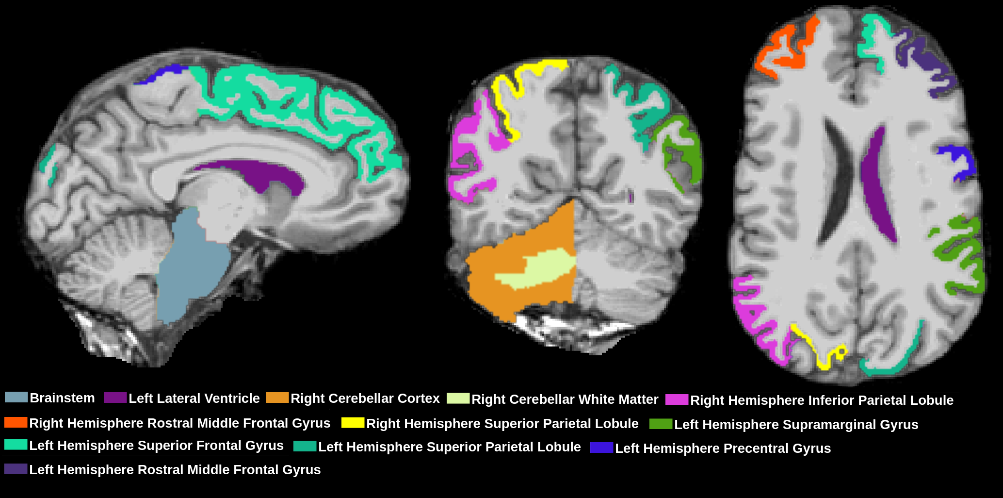
Results: We identified two PD subtypes, differing significantly in structural brain volumes. The regions that most contributed to the differentiation between clusters were the left lateral ventricle, right cerebellar white matter, and the left precentral gyrus.
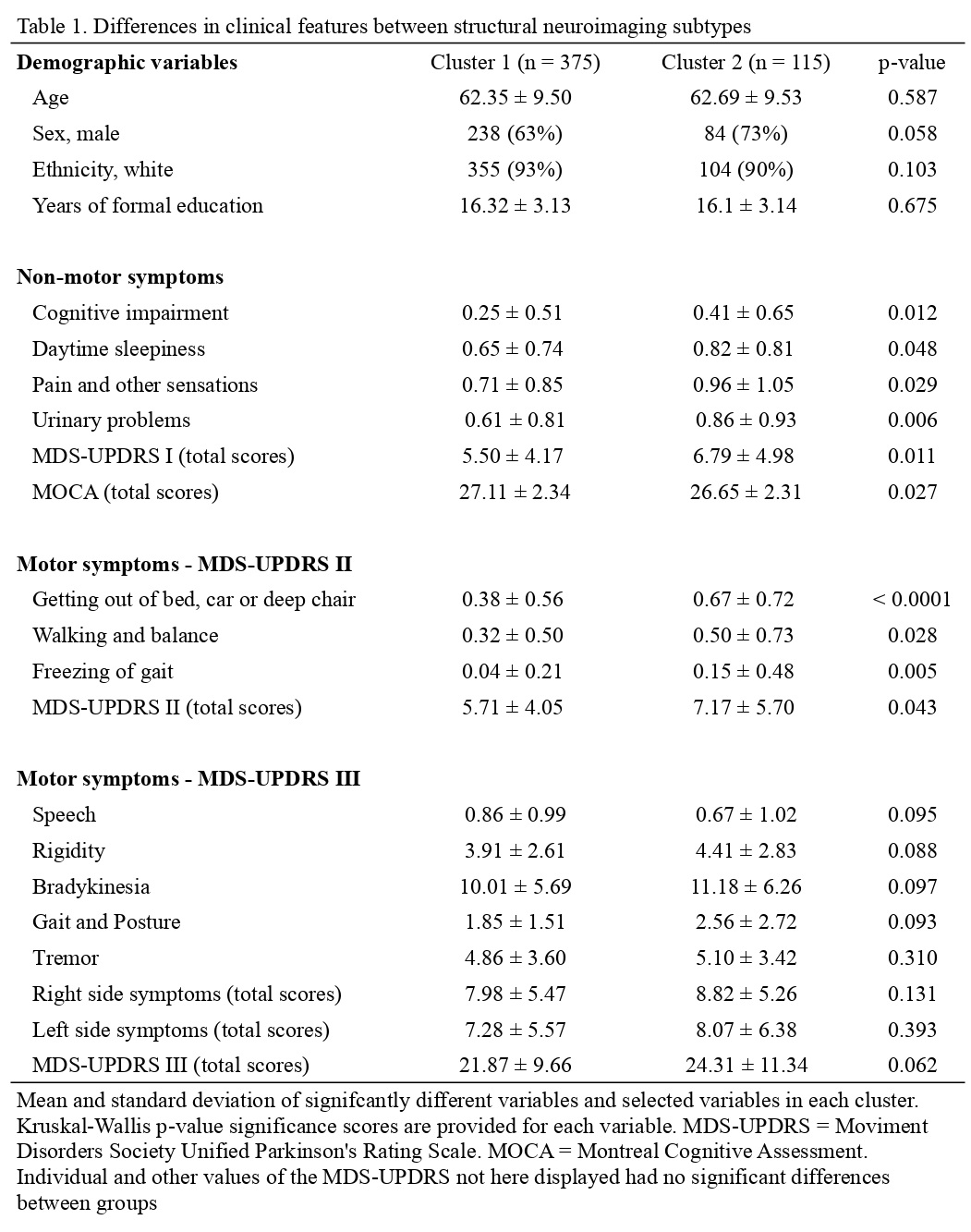
Association analyses, conducted using complete baseline clinical scores, identified two PD subtypes: Cluster 1 (C1; n = 375) with mild symptoms and Cluster 2 (C2; n = 115) with severe symptoms. Both clusters were comparable in age, sex, and education, but C2 showed increased cognitive impairment, daytime sleepiness, pain, urinary problems, and specific axial symptoms like transfer difficulties, walking, balance, and freezing of gait. A slight difference in total MOCA scores was also noted.
Conclusion: We identified two PD subtypes using structural neuroimaging, mainly differentiated by changes in the left lateral ventricle, right cerebellar white matter, and left precentral gyrus. These subtypes showed unique clinical characteristics that are not attributable to demographic differences between the clusters.
Regions included in the cluster analyses
Clinical differences between clusters
To cite this abstract in AMA style:
D. Teixeira-Dos-Santos, A. De-Oliveira-Franco, A. Bieger, C. Mattjie, F. Suzuki, G. Magalhães Pereira, J. Rossi Catao, L. Angi Souza, L. Silveira Kupssinskü, L. Vinícius Moura, MA. Machado Schlindwein, R. Ravazio, S. Duarte Pinto, T. Hugentobler Schlickmann, E. R. Zimmer, MA. de Bastiani, R. C. Barros, AF. Schumacher Schuh. From Images to Insights: Subtyping Parkinson’s Disease Using Unsupervised Learning on MRI Data [abstract]. Mov Disord. 2024; 39 (suppl 1). https://www.mdsabstracts.org/abstract/from-images-to-insights-subtyping-parkinsons-disease-using-unsupervised-learning-on-mri-data/. Accessed November 21, 2025.« Back to 2024 International Congress
MDS Abstracts - https://www.mdsabstracts.org/abstract/from-images-to-insights-subtyping-parkinsons-disease-using-unsupervised-learning-on-mri-data/


