Category: Surgical Therapy: Parkinson's Disease
Objective: To describe the topography of unilateral subthalamic nucleus (STN) lesions performed by magnetic-resonance guided focused ultrasound (MRgFUS) in Parkinson’s disease (PD), and define correlations between lesion size/location and improvement of cardinal signs.
Background: Development of MRgFUS as a minimally invasive approach for lesioning deep brain structures has revitalized ablation as a therapeutic approach for movement disorders. Recently, a pilot study by Martínez-Fernández et al. [1] showed safety and preliminary efficacy of MRgFUS unilateral subthalamotomy for the treatment of all cardinal PD motor features.
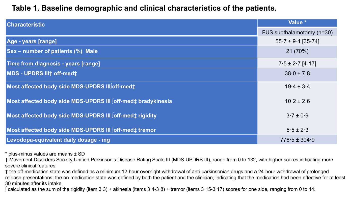
Method: We analyzed volume and location of STN lesions in the T1/T2 MR(3T) images at 24-hours post-procedure in 30 PD patients treated unilaterally (see [Table 1] for baseline characteristics). Lesion topography was correlated to changes in the motor MDS-UPDRS from baseline to 4-months follow-up. MDS-UPDRS-III was evaluated in “off” medication, after 24-hours of drug withdrawal, and sub-items for bradykinesia, rigidity and tremor from body side contralateral to subthalamotomy were scored and analyzed independently. Improvements of ≥60% for each specific motor sign were considered as therapeutic and those with ≤30% as sub-therapeutic, defining two groups for analysis (responders vs non-responders). Lesions providing moderate improvement (30%-60%) were excluded from analysis.
Results: Baseline MDS-UPDRS-III off-state was 38.0±7.0 with a score of 19.4±3.4 corresponding to the most affected (treated) side. Improvement on the treated side was 55.3±19.4%. Change from baseline (%) was 56 ±29.3; 48±26.1 and 67.3±32.1 for rigidity, bradykinesia and tremor respectively. Averaged center lesion in the post-treatment MRI was located within the dorso-lateral STN motor region with a mean volume of 303±127 mm3.
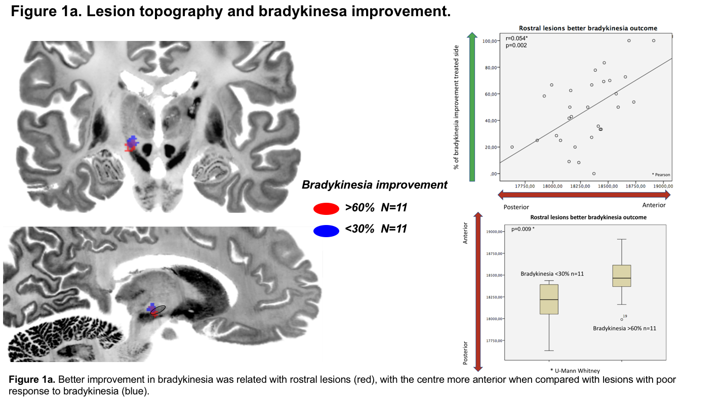
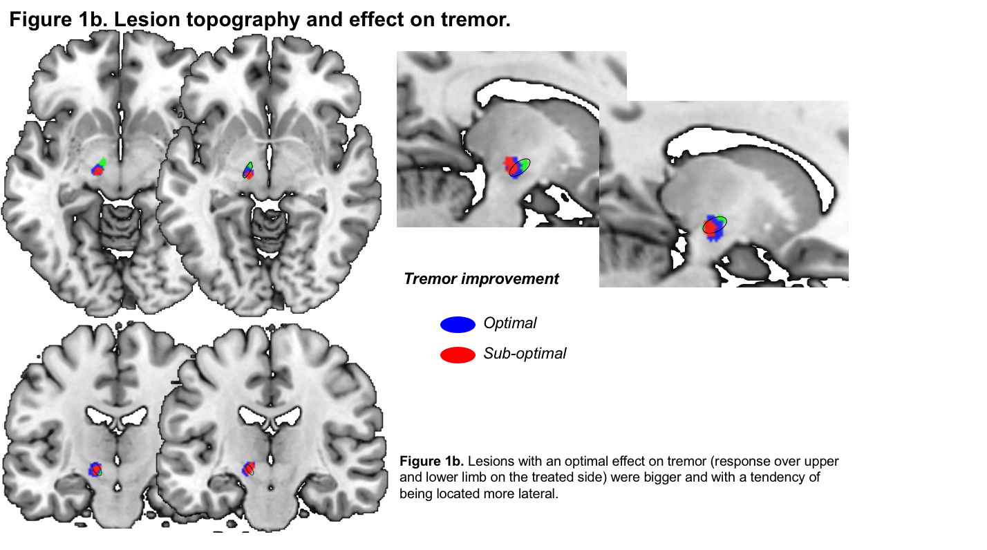
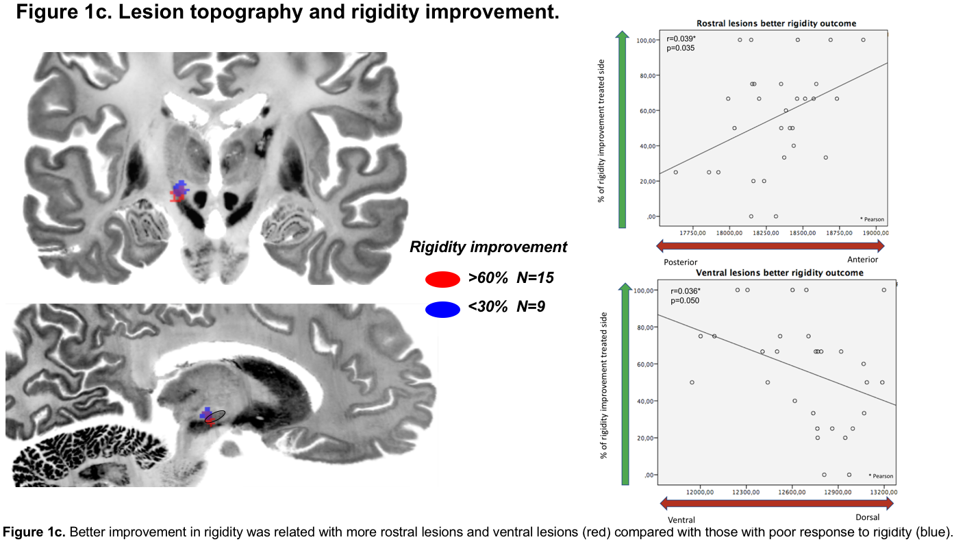
STN lesions with the greatest impact on bradykinesia were significantly more anterior [Figure 1a] than those improving tremor, which lay more laterally; [Figure 1b] rigidity reduction was related to anterior and ventral lesions. [Figure 1c]
Conclusion: Improvement in cardinal motor features after unilateral MRgFUS subthalamotomy is related to specific lesion topography within the motor STN, i.e., bradykinesia rostrally, tremor laterally and rigidity ventro-rostrally. This suggests a segregation within the STN motor subregion for each specific motor feature.
References: 1. Martínez-Fernández R, Rodríguez-Rojas R, Del Álamo M, et al. Focused ultrasound subthalamotomy in patients with asymmetric Parkinson’s disease: a pilot study. Lancet Neurol. 2018;17(1):54–63. doi:10.1016/S1474-4422(17)30403-9.
To cite this abstract in AMA style:
J. Máñez-Miró, R. Rodríguez-Rojas, M. del Álamo, J.A Pineda-Pardo, F. Hernandez-Fernandez, M. Monje, B. Fernandez Rodriguez, C. Gasca-Salas, M. Matarazzo, F. Alonso-Frech, L. Vela-Desojo, R. Martinez-Fernandez, J. Obeso. Focused ultrasound subthalamotomy in Parkinson’s disease: Lesion topography and improvement in cardinal motor features [abstract]. Mov Disord. 2020; 35 (suppl 1). https://www.mdsabstracts.org/abstract/focused-ultrasound-subthalamotomy-in-parkinsons-disease-lesion-topography-and-improvement-in-cardinal-motor-features/. Accessed April 26, 2025.« Back to MDS Virtual Congress 2020
MDS Abstracts - https://www.mdsabstracts.org/abstract/focused-ultrasound-subthalamotomy-in-parkinsons-disease-lesion-topography-and-improvement-in-cardinal-motor-features/


