Session Information
Date: Sunday, October 7, 2018
Session Title: Technology
Session Time: 1:45pm-3:15pm
Location: Hall 3FG
Objective: To review advances in the analysis of the characteristics and pathophysiology of facial movement abnormalities in PD.
Background: Hypomimia, a reduction or absence of spontaneous facial movement and emotional facial expression, is a distinctive clinical manifestation of not only Parkinson’s disease (PD), but also other atypical parkinsonian disorders. Minor variations in the expression of this clinical sign could be useful to differentiate PD from its mimics.
Methods: Medline on PubMed database was searched to identify relevant articles published up to Febuary 2018 as well as manual search. 44 studies related to facial movement in PD were reviewed to explore of characters, pathophysiology and methods and devices of analysis system.
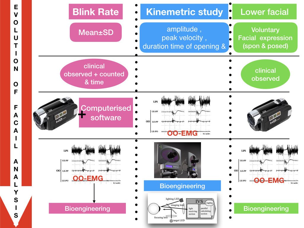
Results: For the last few decades, the clinical observation of facial movement abnormalities in PD was limited to a reduction in spontaneous and emotional facial expression that improved after dopaminergic medications or deep brain stimulation, with the UPDRS-III used for severity grading. With the advent of analytical studies, initially video analysis and subsequently by computerised software, demonstrated a 30-50% reduction in spontaneous blink rate especially while reading, and a reduction in spontaneous perioral movement and emotional facial expression. Neurophysiological studies with OO-EMG further expanded understanding of the reductions in spontaneous blink rate, demonstrating that PD patients had normal voluntary blinking in terms of amplitude and velocity, but a prolonged pause between the closing and opening phase. Voluntary orobuccal movement was also impaired, with smaller amplitude and slower velocity compatible with facial bradykinesia, and that the reduction in emotional facial expression, with both posed and voluntary emotion, is related to the recognition process and cognitive impairment. Recently, sophisticated bioengineering technologies, including magnetic search coil techniques, motion analyzer systems and, high speed blinking analysis systems have demonstrated more detailed abnormalities such as a spontaneous small blink wave prior to blink onset in PD patients.
Conclusions: There has been an evolution in technologies that can analyse facial movement in PD patients enabling a better description of their characteristics and pathophysiology which is helpful when differentiating between parkinsonism disorders.
References: -Bentivoglio AR, Bressman SB, Cassetta E, Carretta D, Tonali D, and Albanese A. Analysis of Blink Rate Patterns in Normal Subjects. Mov DIsor 1997;12: 1028-34. -Deuschl G, Goddemeier C. Spontaneous and reflex activity of facial muscles in dystonia, Parkinson’s disease, and in normal subjects. J Neurol Neurosurg Psychiatry 1998;64:320–4. -Bologna M, Fasano A, Modugno N, et al. Effects of subthalamic nucleus deep brain stimulation and L-dopa on voluntary, spontaneous and reflex blinking in Parkinson’s disease. Exp Neurol 2012;235:265–72. Katsikitis M, Pilowsky I. A study of facial expression in Parkinson’s disease using a novel microcomputer-based method. J Neurol Neurosurg Psychiatry 1988;51:362–6. Hamedani AG and Gold DR. Eyelid Dysfunction in Neurodegenerative, Neurogenetic, and Neurometabolic Disease.Front Neurol 2017;8.
To cite this abstract in AMA style:
W. Rattanachaisit, R. Bhidayasiri. Facial Movement Analysis in Parkinson’s disease: A decade of advances [abstract]. Mov Disord. 2018; 33 (suppl 2). https://www.mdsabstracts.org/abstract/facial-movement-analysis-in-parkinsons-disease-a-decade-of-advances/. Accessed December 16, 2025.« Back to 2018 International Congress
MDS Abstracts - https://www.mdsabstracts.org/abstract/facial-movement-analysis-in-parkinsons-disease-a-decade-of-advances/

