Category: Cognitive Disorders (non-PD)
Objective: find out the relationship between eye movements and cognitive function (CF).
Background: working in our clinic we have learned to give a pre-assessment of CF with just a neurological examination and we would like to share this finding with you.
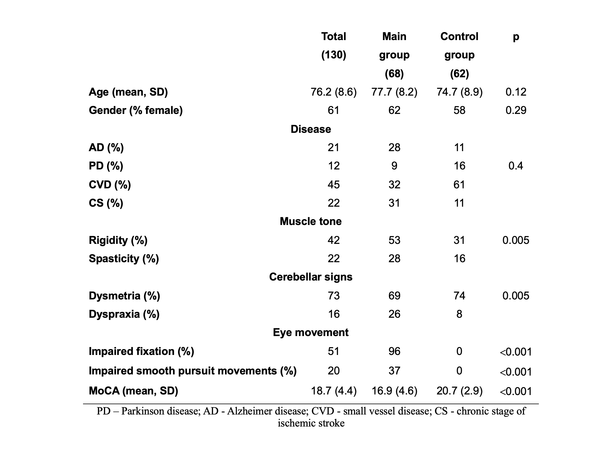
Method: a case-control study were conducted among patients with cerebrovascular and neurodegenerative diseases. The mean age was 76.2 (8.6) and total number of participants was 130. 68 patients of the main group and 62 patients of the control group. The selection criterion for the main group was the presence of impaired smooth pursuit eyes movements (SPEM) and fixation (F). Each patient underwent neurological examination and MoCA test [table1].
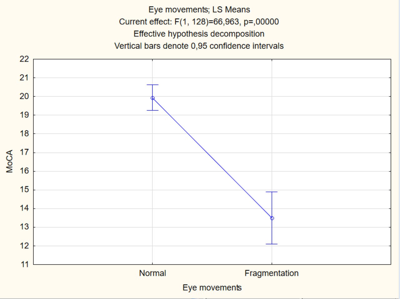
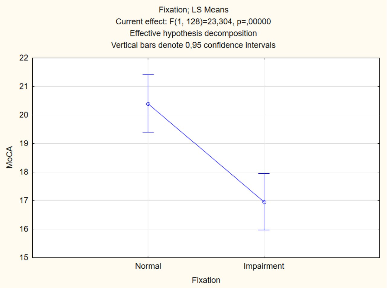
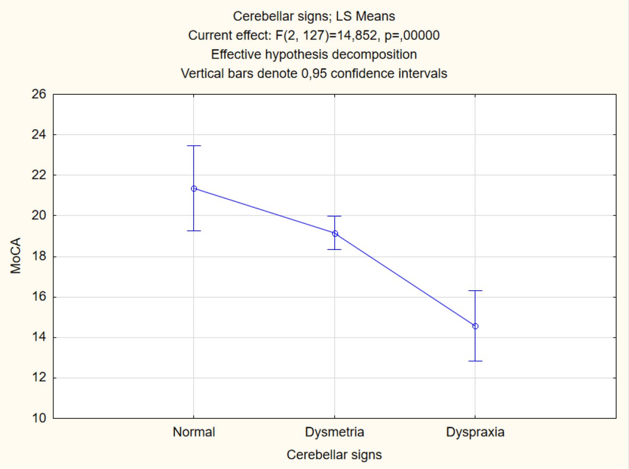
Results: our study revealed that one of the main reflector of CF were eyes (p<0.001), particularly the study of SPEM and F (p<0.001). Also, we found that there was some cerebellar influence on CF (p=0.005). Thus, the present of fragmentation of SPEM was associated with the lowest MoCA score 13.6 (4.3) [figure1]. Presence of dyspraxia of the finger-finger test (FFT) confirmed the presence of dementia (p=0.005). Faulty F negatively affected the results of cognitive tests (р<0.001), but in a less degree than in the case of fragmentation SPEM (MoCA 18.8 (3.7) [figure2]. Most of the patients in the main group had cerebellar signs (mild action tremor or dysmetria) and some had FFT dyspraxia. FFT dyspraxia was associated with a lower MoCA score (р<0.001) [figure3].
By analyzing our findings with anatomophysiological mechanisms [figure4] we came to the following hypotheses [1, 2]:
– Impaired F and altered FFT (more often action tremor) in a group of patients with cognitive decline (MoCA 19 (3.4) may be related to the spread of the pathologic process at the level of the pons structures and cerebellum [1, 2].
– Fragmentation of SPEM and dyspraxia of the FFT (MoCA 11.7 (3.8) may be associated with the spread of the pathologic process to cortical areas and more pronounced lesions of specific cerebellar areas involved in the modulation of eye movements and cognitive processes [1, 2].
Conclusion: a smooth pursuit eye movements assessment can provide a rough estimate of the extent of a patient’s cognitive decline. The pathologic process may initially involve the pons and cerebellum with a gradual spread to cortical structures with a concomitant decrease in the MoCA score [1, 2]. Future studies should investigate the hypothesis of a relationship between eye movement centers and cognitive function.
Baseline characteristic of participants
Cognitive decline and impaired SPEM
Cognitive decline and fixation impairment
Cognitive decline and cerebellar signs
Regulation of smooth pursuit eye movements [1]
References: 1. Leigh, R. J., & Zee, D. S. (2015). The neurology of eye movements. Contemporary Neurology.
2. Schmahmann, J. D. (2019). The cerebellum and cognition. Neuroscience letters, 688, 62-75.
To cite this abstract in AMA style:
A. Ovchynnykova, Y. Trufanov, M. Trishchynska. Eye movements and cognitive function [abstract]. Mov Disord. 2024; 39 (suppl 1). https://www.mdsabstracts.org/abstract/eye-movements-and-cognitive-function/. Accessed May 1, 2025.« Back to 2024 International Congress
MDS Abstracts - https://www.mdsabstracts.org/abstract/eye-movements-and-cognitive-function/


![Regulation of smooth pursuit eye movements [1]](https://www.mdsabstracts.org/wp-content/uploads/2024/09/0863_1287_000245_5.png)