Category: Huntington's Disease
Objective: This study aims to investigate correlations between mHtt and NfL levels and MRI findings in individuals at different stages of HD, including presymptomatic and symptomatic individuals.
Background: Biomarkers in Huntington’s disease (HD) are crucial for understanding disease progression and developing effective therapies. Additionally, cerebrospinal fluid (CSF) biomarkers like neurofilament light chain (NfL) are sensitive indicators of neurodegeneration, while mutant Huntingtin (mHtt) is specific to HD. Magnetic resonance imaging (MRI) provides detailed insights into brain structure and neurodegenerative changes. However, there’s a lack of studies using multimodal techniques to correlate specific brain structures’ neurodegeneration with CSF biomarkers.
Method: Longitudinal study with cross-sectional analysis of a cohort of individuals with genetic confirmation for Huntington’s disease (HD) and controls. They were grouped into presymptomatic (preHD) at the motor level (DLC<4) and symptomatic (HD) (DCL=4). Levels of NfL in CSF (Simoa, Quanterix) and mHTT in CSF (SMC Erenna platform, Merck) were quantified. MRI 3 Tesla and gray matter analysis using VBM and cortical thickness using FreeSurfer were performed. Correlations between NfL and mHtt biomarkers and neuroimaging were established using SPM.
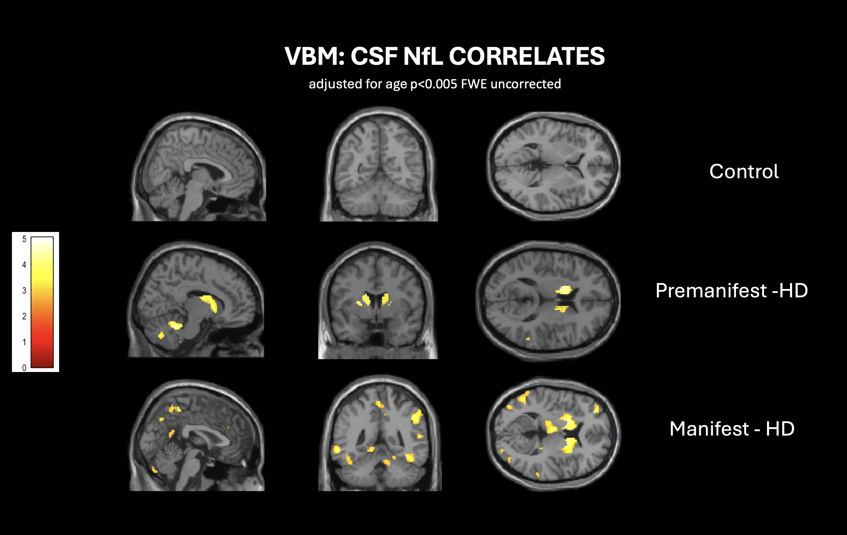
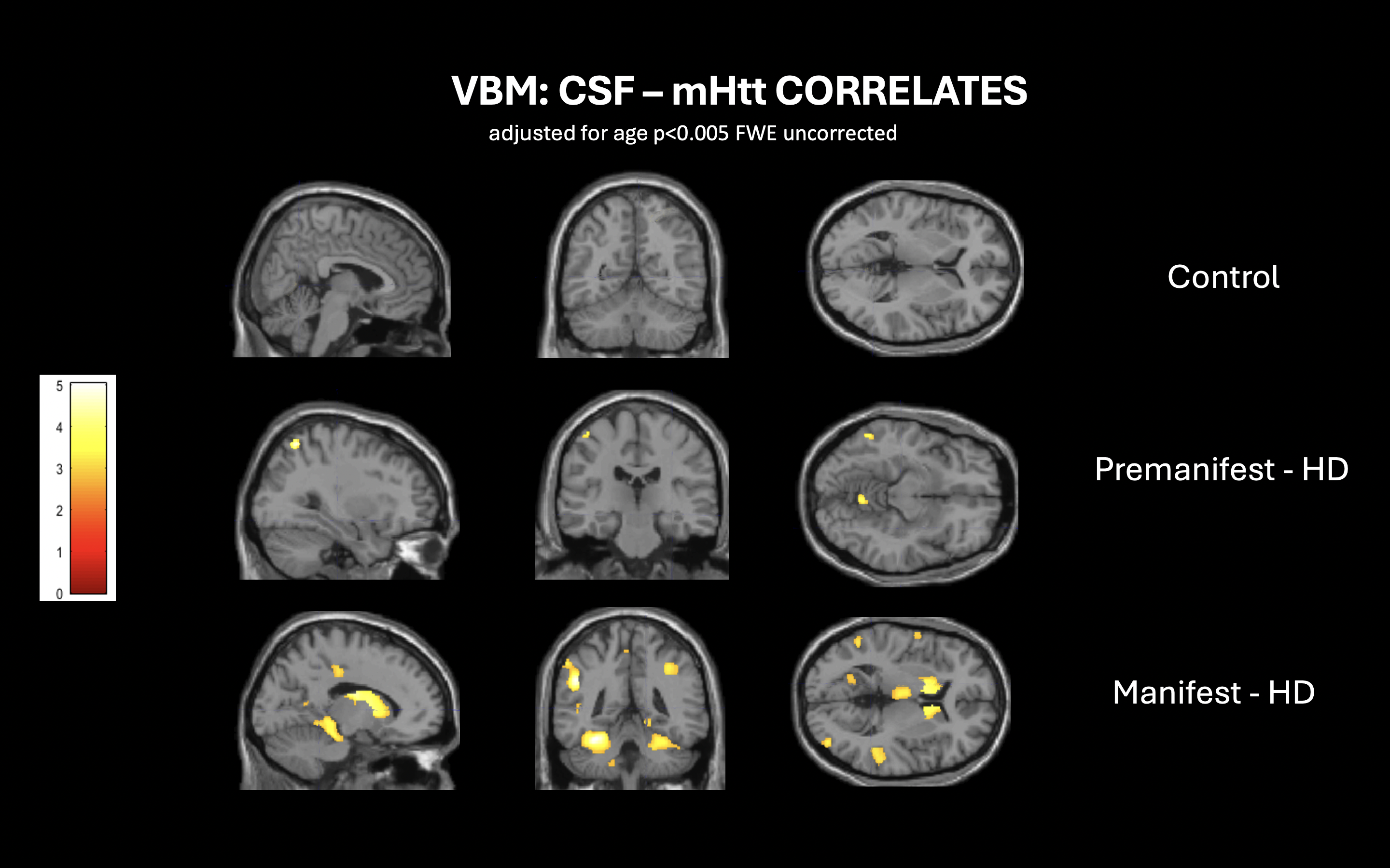
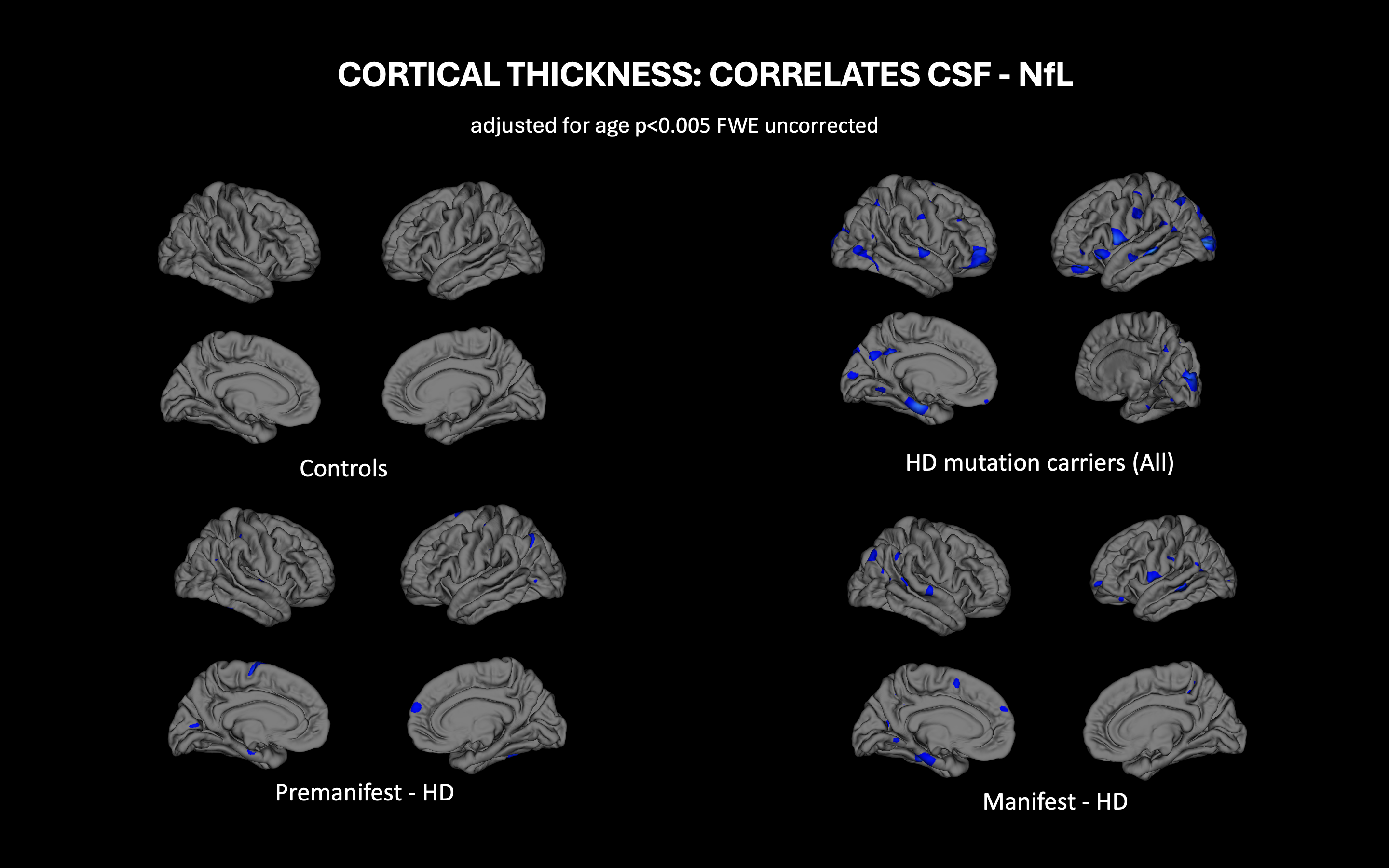
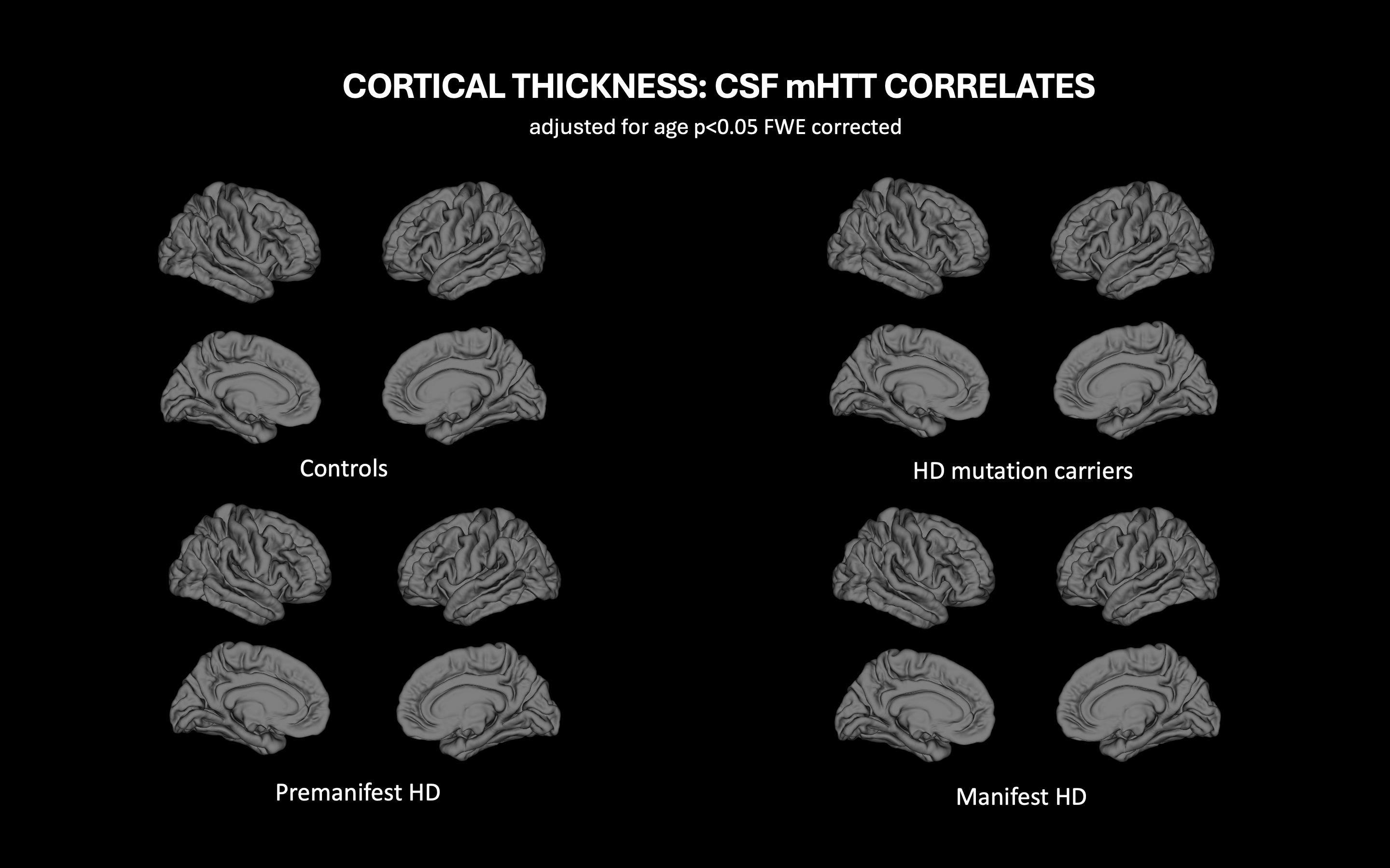
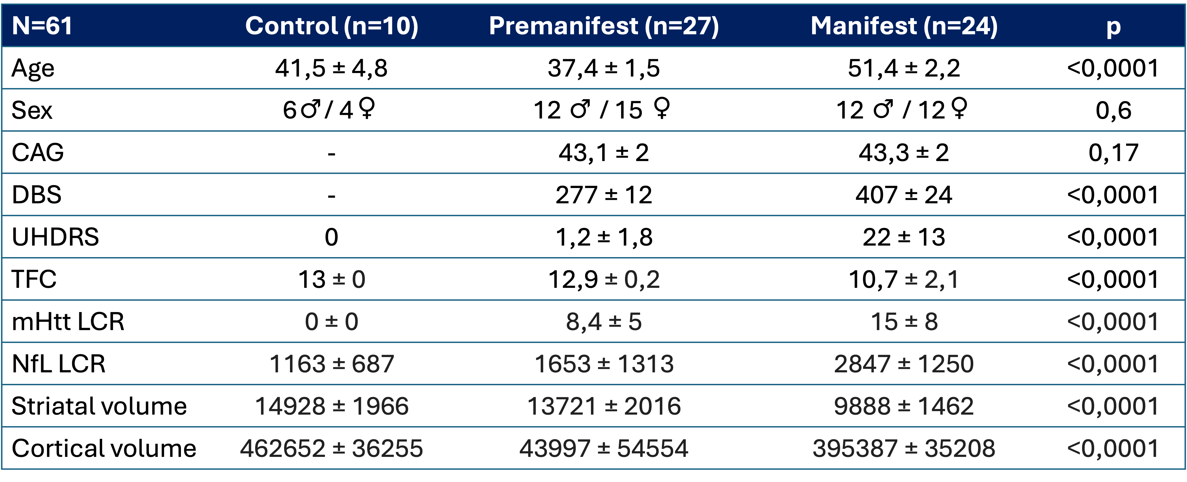
Results: Sixty-one participants, including 27 presymptomatic, 24 symptomatic, and 10 controls, showed age differences between groups. Presymptomatic individuals exhibited elevated NfL and mHtt levels compared to controls, with further increases in symptomatic individuals (table 1). NfL levels correlated with caudate, putamen, and cerebellar atrophy in presymptomatics (p<0.005), while mHtt levels showed weaker correlation (p<0.05). In symptomatics, NfL levels correlated with thalamic, posterior cortical, and prefrontal atrophy, whereas mHtt levels correlated less with posterior cortical atrophy, suggesting NfL higher sensitivity in detecting neurodegeneration and/or involvement of non-Huntingtin-dependent processes (figures 1-4)
Conclusion: Levels of NfL exhibit greater sensitivity in detecting the neurodegenerative process compared to mHtt, showing a correlation with atrophy in the caudate and putamen in presymptomatic individuals that extends to cortical areas as the disease progresses.
Figure 1
Figure 2
Figure 3
Figure 4
Table 1
References: Byrne et al. Neurofilament light protein in blood as a potential biomarker of neurodegeneration in Huntington’s disease: a retrospective cohort análisis Lancet Neurology 2017
Rodriguez F. Biofluid Biomarkers in Huntington’s Disease Huntington’s Disease, Methods in Molecular Biology, 2020
Johnson et al. Neurofilament light protein in blood predicts regional atrophy in Huntington disease, Neurlogy 2018
To cite this abstract in AMA style:
J. Pérez, S. Horta, G. Saura, A. Barba, A. Davi, A. Vázquez, E. Rivas, A. Campolongo, J. Pagonabarraga, J. Kulisevsky. Exploring Correlations between Mutant Huntingtin, NfL, and MRI in Huntington’s Disease: A Multimodal Analysis [abstract]. Mov Disord. 2024; 39 (suppl 1). https://www.mdsabstracts.org/abstract/exploring-correlations-between-mutant-huntingtin-nfl-and-mri-in-huntingtons-disease-a-multimodal-analysis/. Accessed December 23, 2025.« Back to 2024 International Congress
MDS Abstracts - https://www.mdsabstracts.org/abstract/exploring-correlations-between-mutant-huntingtin-nfl-and-mri-in-huntingtons-disease-a-multimodal-analysis/

