Category: Surgical Therapy: Parkinson's Disease
Objective: The objective of the study was to determine the anatomical localisation of evoked resonant neural activity (ERNA), which is being investigated as a potential physiological guidance signal for targeting electrodes in deep brain stimulation (DBS) of the subthalamic nucleus (STN) in Parkinson’s disease (PD).
Background: Deep brain stimulation of the STN is well-established for the treatment for PD with motor fluctuations. Targeting of DBS electrodes is generally performed using a combination of anatomical landmarks, intra-operative microelectrode recording (MER), and clinical stimulation testing. Electrophysiological signals typically used for targeting during MER lack anatomical precision and signal robustness. ERNA offers a robust signal for electrophysiological targeting and is readily detectable during both awake and asleep (general anesthesia) conditions,[1] however its anatomical precision and origin are unknown.
Method: Anatomical neuroimaging and electrophysiological data was collected from 42 patients (75 hemispheres) with PD who underwent routine bilateral DBS of the STN using Medtronic 3387 electrodes. Pre-operative brain MRI and post-operative computed tomography images were co-registered and normalized to MNI (Montreal Neurological Institute) space to determine electrode location. Intraoperative recordings of ERNA were made by applying stimulation and recording the evoked resonant signal. Root mean square (RMS) power of the signal was then calculated and mapped to electrode location to obtain a heatmap for ERNA.
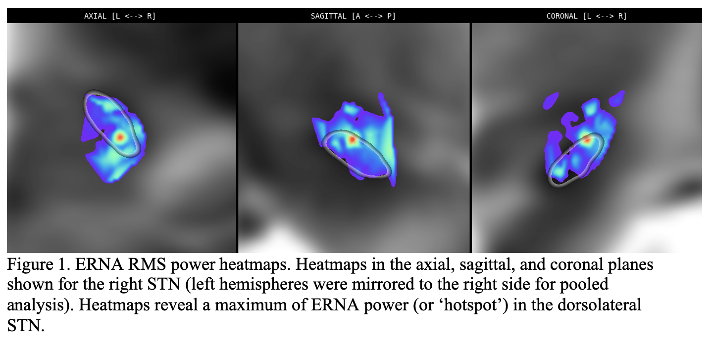
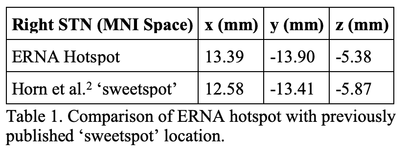
Results: The ERNA RMS power heatmap revealed a maximum in dorsolateral STN, within the region typically targeted for DBS [figure1]. It falls within the motor STN subdivision, close to the border with the associative subdivision. This ERNA ‘hotspot’ is located close to hotspots published using other methods,[2] but slightly lateral and posterior [table1].
Conclusion: ERNA is maximal within the dorsolateral (motor) STN and is close to previously published optimal stimulation target locations. These findings lend weight to the hypothesis that the circuits that generate ERNA form part of the neural circuitry underlying the pathophysiology of parkinsonism. Together with the robust nature of the signal, we propose that ERNA can be used as a reliable and precise biomarker in the targeting of STN DBS electrodes for Parkinson’s disease.
References: 1. Sinclair, N. C. et al. Electrically evoked and spontaneous neural activity in the subthalamic nucleus under general anesthesia. J. Neurosurg. 1, 1–10 (2021).
2. Horn, A., Neumann, W. J., Degen, K., Schneider, G. H. & Kühn, A. A. Toward an electrophysiological “Sweet spot” for deep brain stimulation in the subthalamic nucleus. Hum. Brain Mapp. 38, 3377–3390 (2017).
To cite this abstract in AMA style:
H. Simpson, S. Xu, W. Lee, A. Begg, K. Bulluss, H. Mcdermott, W. Thevathasan, T. Perera. Evoked resonant neural activity within the dorsolateral subthalamic nucleus as a biomarker for the targeting of deep brain stimulation for Parkinson’s disease [abstract]. Mov Disord. 2022; 37 (suppl 2). https://www.mdsabstracts.org/abstract/evoked-resonant-neural-activity-within-the-dorsolateral-subthalamic-nucleus-as-a-biomarker-for-the-targeting-of-deep-brain-stimulation-for-parkinsons-disease/. Accessed January 7, 2026.« Back to 2022 International Congress
MDS Abstracts - https://www.mdsabstracts.org/abstract/evoked-resonant-neural-activity-within-the-dorsolateral-subthalamic-nucleus-as-a-biomarker-for-the-targeting-of-deep-brain-stimulation-for-parkinsons-disease/


