Category: Surgical Therapy: Parkinson's Disease
Objective: Compare the evoked resonant neural activity (ERNA) responses obtained from stimulation of Subthalamic Nucleus (STN) and Globus Pallidus Internus (GPi) in patients with Parkinson’s disease (PD).
Background: ERNA has recently been investigated as a possible biomarker for optimizing lead placement and stimulation parameter selection in deep brain stimulation (DBS) of PD patients [1]. Typically, the main DBS targets for PD are STN or GPi, which are equally effective in controlling the motor symptoms of PD. However, the optimal target for a given patient is still uncertain. The majority of ERNA characterization has been within STN with only a few studies investigating ERNA in GPi and comparing the two responses.
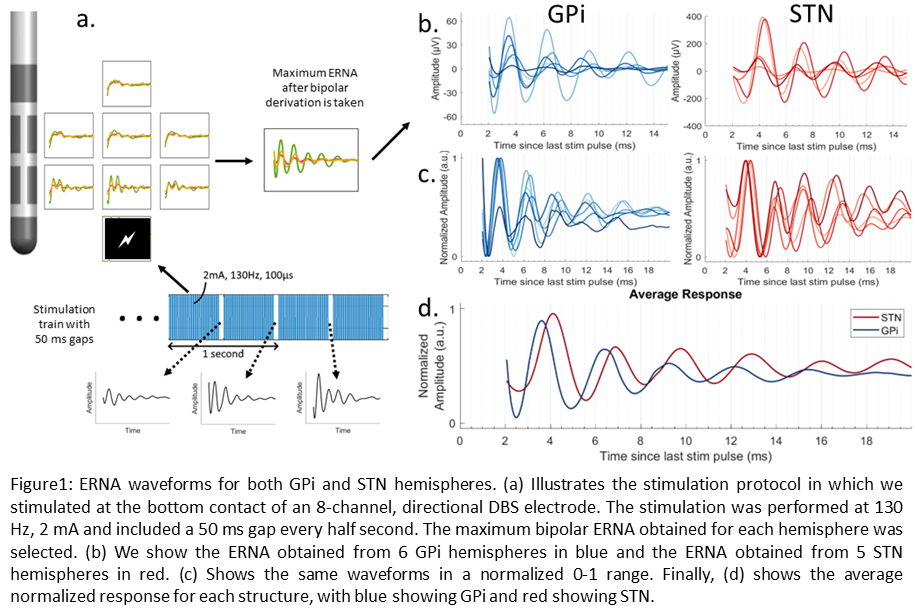
Method: Here we present intraoperative LFP data collected during awake GPi and STN implantations of six PD patients with 8-channel directional electrodes (4 levels). Monopolar simulation was delivered at the bottom contact (Lv1), with 130 Hz, 2mA for 25 seconds (Fig. 1a). Our stimulation train also incorporated a 50ms gap at a rate of 2 gaps per second for a total of 50 ERNA waveforms per hemisphere. An average ERNA waveform was computed for all contacts expect Lv1 due to stimulation artifact. After taking bipolar derivations, the largest ERNA waveforms were selected for each hemisphere. One STN response was excluded as no ERNA was detected, possibly due electrode placement. After normalizing each waveform, we also computed the average ERNA waveform across GPi and STN structures.
Results: The first peak of GPi-ERNA had an average amplitude of 16.5 µV with ±17.3 µV standard deviation while the first peak of STN-ERNA was, on average, 90.5 µV with ±90.6 µV standard deviation as shown in Fig. 1b. Consistently, the GPi-ERNA had a smaller latency than STN-ERNA producing a first peak after the last stim pulse at around 3.6 ms for GPi and 4.1 ms for STN (Fig. 1d), in agreement with previous studies [2]. The ERNA from GPi also decayed faster compared to STN (Fig. 1b-d).
Conclusion: We have demonstrated ERNA in both STN and GPi patients with PD. Our results show that ERNA amplitudes are smaller in GPi compared to STN. Moreover, ERNA latency is consistently smaller in GPi compared to STN.
References: 1- Ozturk, Musa, et al. “Electroceutically induced subthalamic high-frequency oscillations and evoked compound activity may explain the mechanism of therapeutic stimulation in Parkinson’s disease.” Communications Biology 4.1 (2021): 393.
2- Schmidt, Stephen L., et al. “Evoked potentials reveal neural circuits engaged by human deep brain stimulation.” Brain stimulation 13.6 (2020): 1706-1718.
To cite this abstract in AMA style:
L. Branco, C. Swamy, A. Tarakad, N. Vanegas-Arroyave, I. Patel, N. Ince, A. Viswanathan. Evoked resonant neural activity in Globus Pallidus vs. Subthalamic Nucleus of patients with Parkinson’s Disease, preliminary results [abstract]. Mov Disord. 2023; 38 (suppl 1). https://www.mdsabstracts.org/abstract/evoked-resonant-neural-activity-in-globus-pallidus-vs-subthalamic-nucleus-of-patients-with-parkinsons-disease-preliminary-results/. Accessed January 2, 2026.« Back to 2023 International Congress
MDS Abstracts - https://www.mdsabstracts.org/abstract/evoked-resonant-neural-activity-in-globus-pallidus-vs-subthalamic-nucleus-of-patients-with-parkinsons-disease-preliminary-results/

