Category: Rare Genetic and Metabolic Diseases
Objective: We aim to study the sensitivity and specificity of hyperintense globus pallidus rim sign in WD diagnosis.
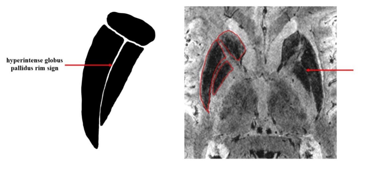
Background: The diagnosis of Wilson disease (WD) remains challenging due to the wide range of phenotypic manifestations with overlapping ages of presentation and multisystem involvement. Although brain MRI are also included in the diagnostic approach, the current MRI features of WD are neither sensitive nor specific. Recently, with susceptibility weighted imaging (SWI) in 7T MRI, we observed a linear signal intensity change consisting of a lateral hyperintense rim at the lateral border of globus pallidus in WD patients (Figure 1). We term the intensity change as hyperintense globus pallidus rim sign.
Method: A total of 35 WD patients, 15 first-degree relatives of WD patients, 18 age-matched health controls and 15 young-onset Parkinson’s disease (YOPD) were enrolled in this study. WD patients were divided into the WD with neurological symptoms (NWD) group and the WD without neurological symptoms (nNWD) group, according to the presence or absence of neurological dysfunctions during the whole clinical course. All participants were scanned in seven-tesla MRI and high spatial resolution SWI were collected. Two neurologists were invited to describe whether hyperintense globus pallidus rim sign is visible in targeted images of basal ganglia and rated the scores of the sign.
Results: In 7T MRI, the hyperintense globus pallidus rim sign was detected in 26 of the 28 NWD patients, and was negative in all 7 nNWD patients, heterozygote ATP7B mutation carriers, PD patients and HC. The diagnostic sensitivity was 92.86% and specificity was 100%, which was significantly higher than the sensitivity and specificity of traditional MRI features of WD. After adjusted for age, disease duration and treatment duration, we found positive correlation between the hyperintense globus pallidus rim sign scores and UWDRS in the NWD group (R=0.448, P=0.025). The hyperintense globus pallidus rim sign was negative in all 15 YOPD patients.
Conclusion: Our study suggested that hyperintense globus pallidus rim sign is an effective neuroimaging biomarker for the diagnosis and differential diagnosis of NWD with high sensitivity and excellent specificity in 7T MRI. It may indicate a special iron deposition pattern which spared the external medullary lamina in NWD patients.
References: [1] Prashanth L, Sinha S, Taly A, Vasudev M. Do MRI features distinguish Wilson’s disease from other early onset extrapyramidal disorders? An analysis of 100 cases. Movement disorders : official journal of the Movement Disorder Society 2010;25(6):672-678. doi: 10.1002/mds.22689
To cite this abstract in AMA style:
D. Su, Z. Zhang, Z. Zhang, T. Wu, J. Jing, T. Feng. Evaluation of hyperintense globus pallidus rim sign in seven-tesla MRI as a diagnostic biomarker in Wilson’s disease [abstract]. Mov Disord. 2022; 37 (suppl 2). https://www.mdsabstracts.org/abstract/evaluation-of-hyperintense-globus-pallidus-rim-sign-in-seven-tesla-mri-as-a-diagnostic-biomarker-in-wilsons-disease/. Accessed December 23, 2025.« Back to 2022 International Congress
MDS Abstracts - https://www.mdsabstracts.org/abstract/evaluation-of-hyperintense-globus-pallidus-rim-sign-in-seven-tesla-mri-as-a-diagnostic-biomarker-in-wilsons-disease/

