Category: Dystonia: Pathophysiology, Imaging
Objective: This study aimed to investigate abnormal cerebellar iron distribution in patients with idiopathic CD using quantitative susceptibility mapping (QSM).
Background: Idiopathic CD is a rare neurologic disorder characterized by involuntary muscle contractions in the neck. Recent neuroimaging investigations demonstrated the association between dystonia and alterations of cerebellar structure, connectivity, and functional activity.
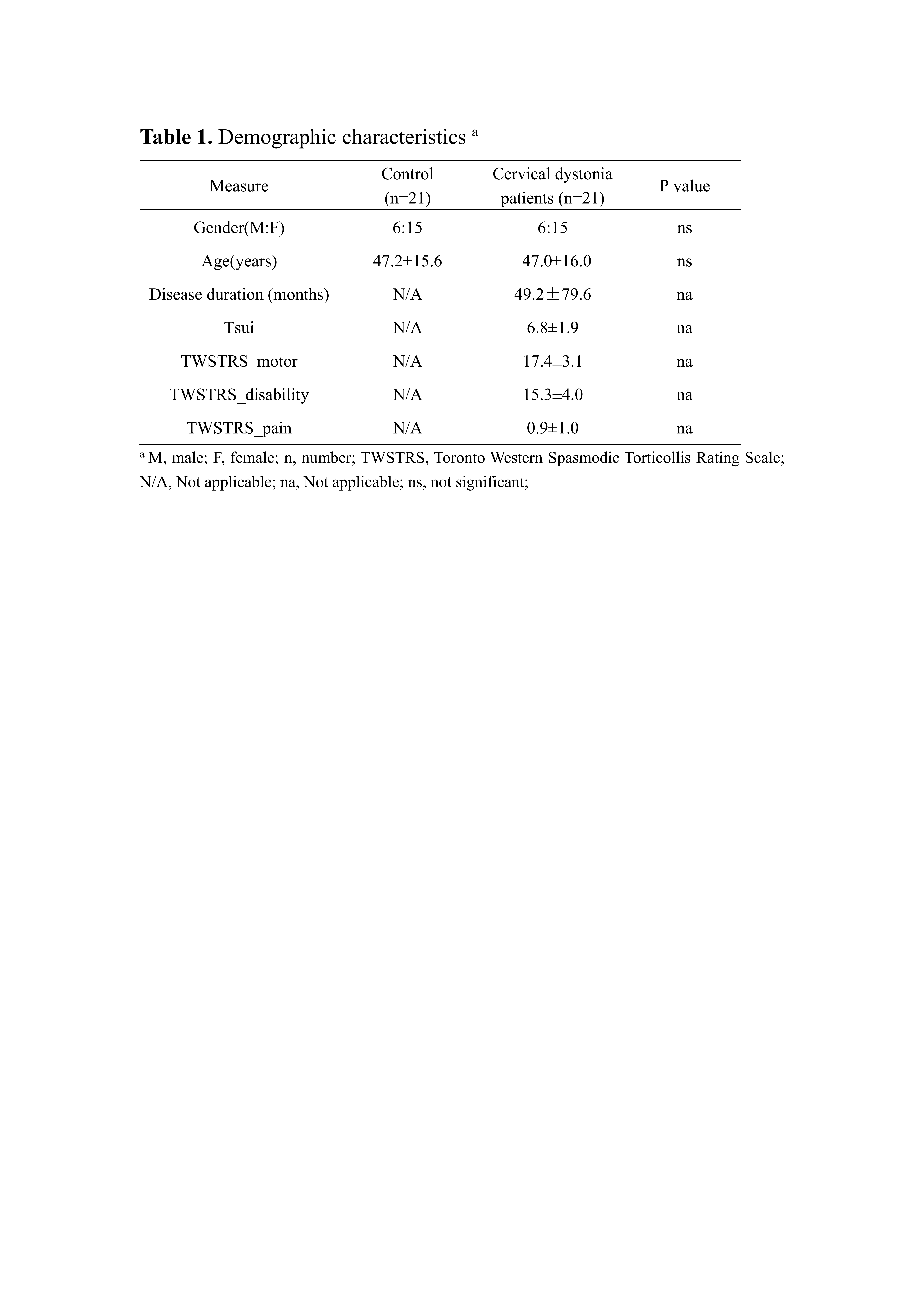
Method: We assessed 21 patients with cervical dystonia (15 females) and age- and sex-comparable healthy control using the Toronto Western Spasmodic Torticollis Rating Scale and Tsui Rating Scale (Table 1). The volumetric alterations of whole-brain gray matter (GM) were evaluated using two voxel-based approaches (SPM-VBM and FSL-VBM). The QSM images were reconstructed from the data acquired using a 3D gradient recalled echo sequence. The voxel-wise analysis was performed to access the brain tissue magnetic susceptibility differences between groups. Regional QSM analyses of the cerebellum were further conducted based on the brain parcellation defined by AAL3 atlas.
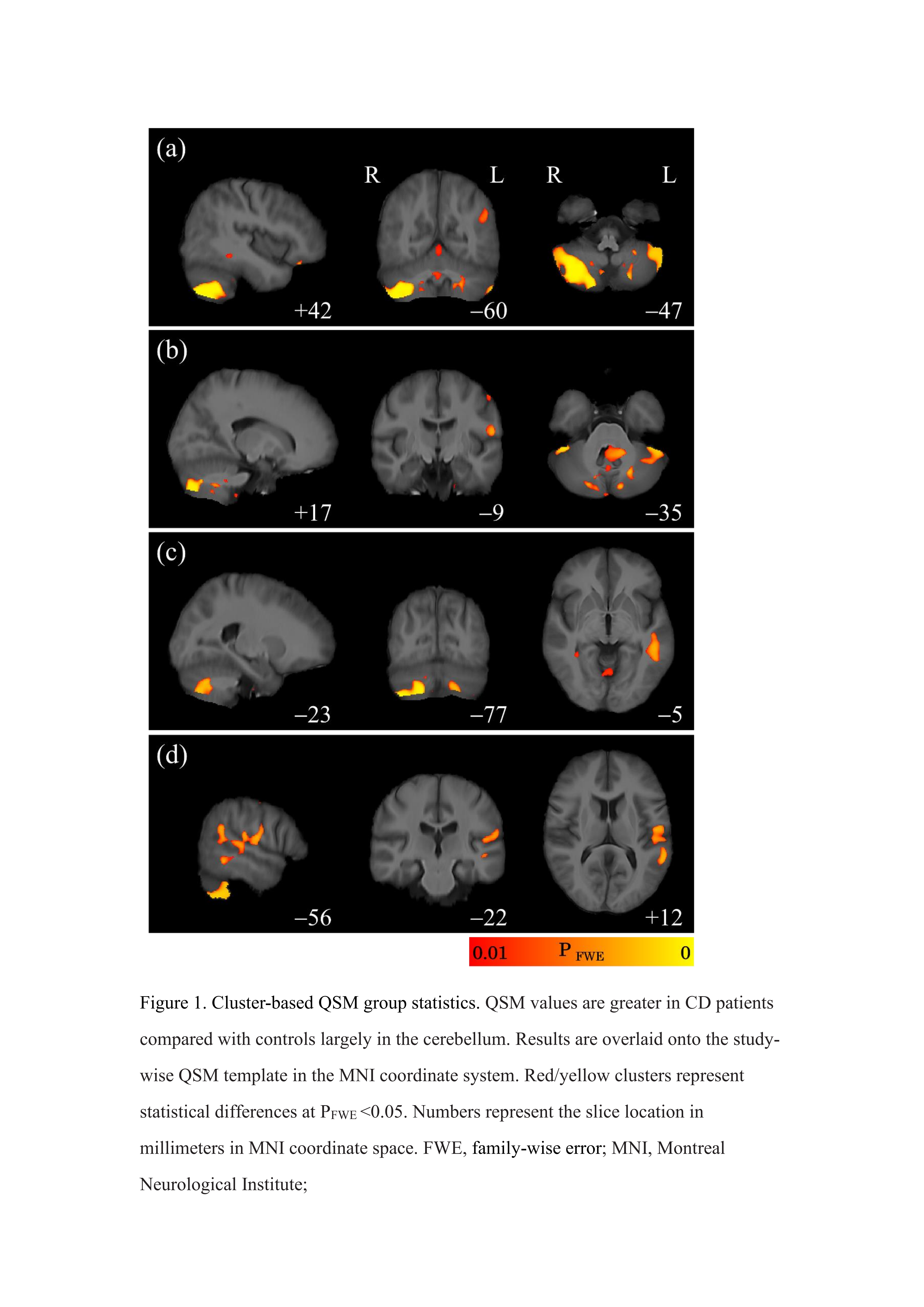
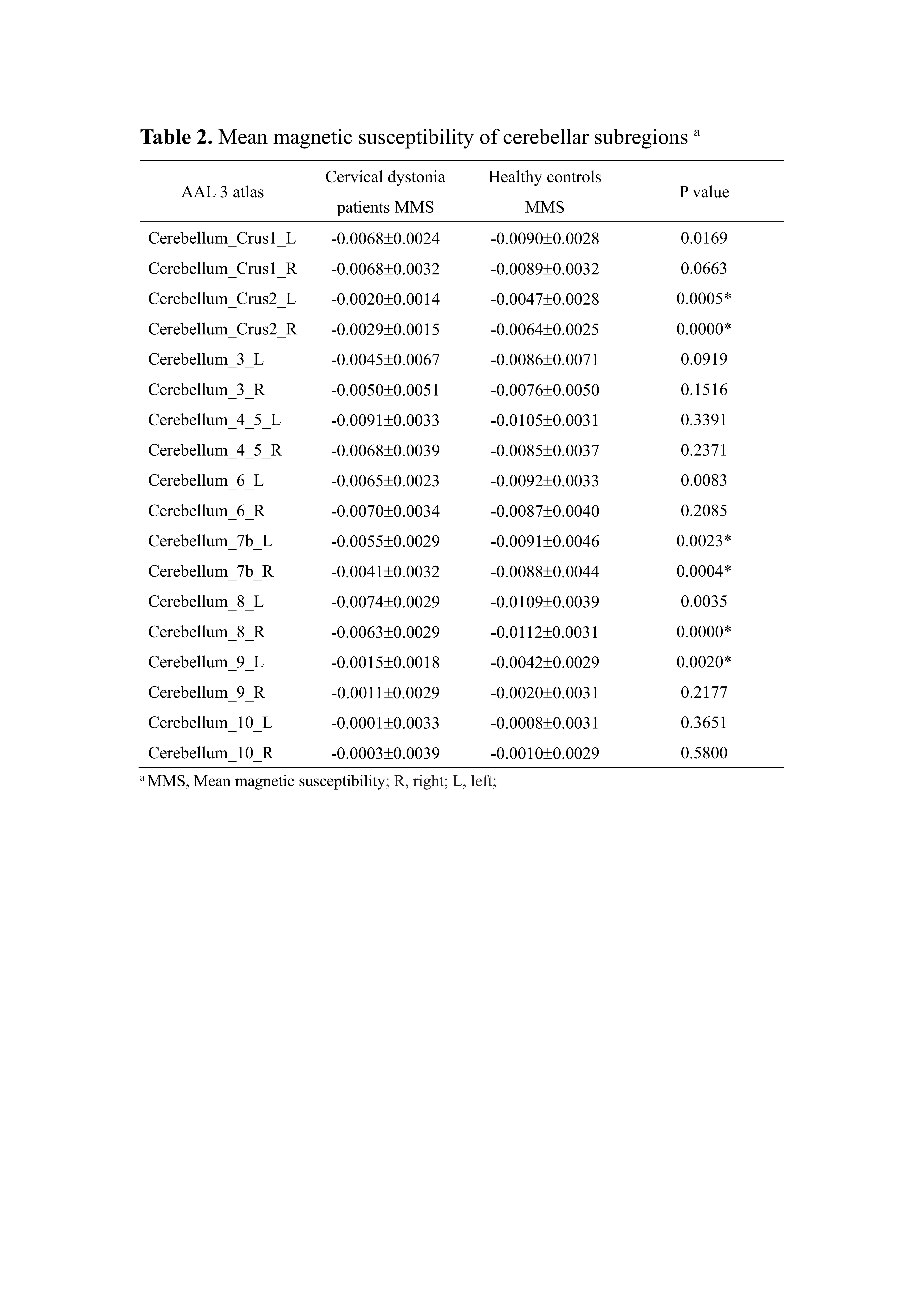
Results: There was no significant difference between groups regarding the whole-brain GM volume (p > 0.05, family-wise error corrected). Interestingly, we found widespread susceptibility increases (p < 0.01, family-wise error corrected) in cervical dystonia compared with healthy controls that primarily involved cerebellar structures, as shown in Figure 1. The regional QSM analyses showed that the susceptibility values were significantly higher in Crus2, lobule ⅦB, right lobule Ⅷ, and left lobule Ⅸ, indicating brain iron accumulation in these regions (p < 0.0028, Bonferroni corrected) (Table 2). However, no significant correlations were found between susceptibility measurement and disease duration or motor severity.
Conclusion: Our study revealed a spatial distribution of QSM alterations in CD patients. Our results showed that iron overload mainly occurred in the Crus2, lobule ⅦB, lobule Ⅷ, which are related to the second-order motor and non-motor processing.In contrast to QSM, no significant effects were observed with structural MRI measurements. These results demonstrate the relevance of mapping the biochemical environment of the CD in vivo, and provide new insights into the behavior of QSM as a disease biomarker for CD.
To cite this abstract in AMA style:
HX. Li, M. Zhang, YH. Wu, YF. Li, HJ. Wei, YW. Wu. Evaluation of abnormal cerebellar iron distribution in idiopathic cervical dystonia using quantitative susceptibility mapping [abstract]. Mov Disord. 2021; 36 (suppl 1). https://www.mdsabstracts.org/abstract/evaluation-of-abnormal-cerebellar-iron-distribution-in-idiopathic-cervical-dystonia-using-quantitative-susceptibility-mapping/. Accessed April 22, 2025.« Back to MDS Virtual Congress 2021
MDS Abstracts - https://www.mdsabstracts.org/abstract/evaluation-of-abnormal-cerebellar-iron-distribution-in-idiopathic-cervical-dystonia-using-quantitative-susceptibility-mapping/


