Session Information
Date: Monday, October 8, 2018
Session Title: Parkinson's Disease: Neuroimaging And Neurophysiology
Session Time: 1:15pm-2:45pm
Location: Hall 3FG
Objective: Neurotropic drugs may modulate Electroencephalography (EEG) microstates (MS), but there are no works showing EEG MS default-mode network changes in response to dopaminergic stimulation in Parkinson disease (PD) patients.
Background: Dopaminergic stimulation is responsible for changes in motor and non-motor symptoms in PD.EEG records the electrical field produced by the brain electrical activity and can be studied trough Microstate (MS) analysis. MSs are defined by topographies of electrical potential recorded in a multichannel matrix, which remain stable for 80-120 ms before rapidly moving to a different MS. MS analysis simultaneously evaluates the signal from all the electrodes to create a global representation of a functional state. EEG MS have been related to fMRI resting state networks.
Methods: 14 PD subjects in HY stage III or less with no clinical evident fluctuations were included, (mean disease duration 6,5± 1,74 years). All patients were receiving dopaminergic stimulation with levodopa or dopaminergic agonists (576 ± 475,04 levodopa equivalents). Resting EEG activity was recorded before the first daily PD medication dose, during two minutes from 64 electrodes placed according to the 10-20 setting. After one hour from drug intake resting EEG activity was again recorded. MATLAB and LORETA software were used for signal processing and characterization. Time and frequency variables for each MS were calculated. The difference of averages between pre and post conditions for each feature was checked by a t-test for repeated measures with bootstrapping (n=2000).
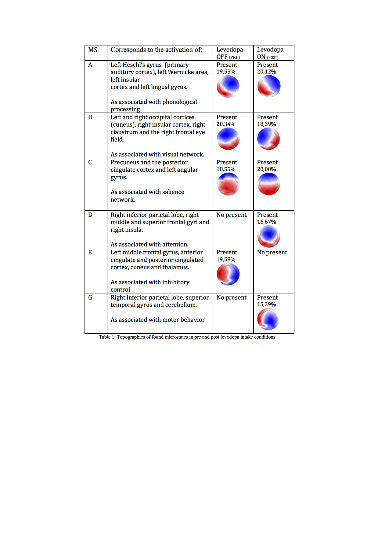
Results: Typical variances of pre and post medication intake in MS are sown in Table 1. MS A duration decreases after levodopa intake, MS B appears more times than before levodopa intake.
Conclusions: The described MS changes have previously been correlated with cognitive fatigue, attention and executive function changes. These changes are cognitive symptoms related to PD. This work demonstrates that there is a change of the EEG MS in PD induced by an increase in dopaminergic stimulation. This neurophysiological test can be useful to characterize pharmacological response of typical PD patients or even detect non-motor fluctuations even before they become evident. This opens a new field for the characterization of the disease. Further studies are required on atypical PD patients.
References: Custo, Anna, Dimitri Van De Ville, William M. Wells, et al. 2017 Electroencephalographic Resting-State Networks: Source Localization of Microstates. Brain Connectivity 7(10): 671-682. Michel, Christoph M., and Thomas Koenig 2017 EEG Microstates as a Tool for Studying the Temporal Dynamics of Whole-Brain Neuronal Networks: A Review. NeuroImage.
To cite this abstract in AMA style:
V. Cortés, I. Serrano, N. Mendes, M. Del Castillo, A. Arroyo, J. Andreo, M. Del Valle, J. Herreros, JP. Romero,. EEG microstates changes in response to increase in dopaminergic stimulation in treated typical Parkinson’s disease patients without clinical fluctuations [abstract]. Mov Disord. 2018; 33 (suppl 2). https://www.mdsabstracts.org/abstract/eeg-microstates-changes-in-response-to-increase-in-dopaminergic-stimulation-in-treated-typical-parkinsons-disease-patients-without-clinical-fluctuations/. Accessed December 20, 2025.« Back to 2018 International Congress
MDS Abstracts - https://www.mdsabstracts.org/abstract/eeg-microstates-changes-in-response-to-increase-in-dopaminergic-stimulation-in-treated-typical-parkinsons-disease-patients-without-clinical-fluctuations/

