Category: Parkinson's Disease: Neuroimaging
Objective: In this study, we aimed to comprehensively investigate the early neurotransmitters changes in a cohort of de novo Parkinson’s disease (PD) patients with and without REM sleep behavior disorder (RBD) by analysing the relationship between regional gray matter volume differences with atlas-based distribution of multiple neurotransmitters.
Background: Uncertainty surrounding the pathology and disruption of neurotransmitter systems in RBD makes it challenging to treat. Only few PET studies have evaluated RBD using specific radiotracers for neurotransmitters and neuroreceptors[1]. Therefore, multisystem neuroimaging methods could help establish the neurochemical features of RBD.
Method: PD patients were selected from the Parkinson’s Progression Markers Initiative cohort who underwent structural MRI. Demographic characteristics and clinical features including RBD screening questionnaire (RBDSQ), were extracted for analyses. Probable RBD was screened on the basis of the RBDSQ with a cut-off score of 6[2]. PD patients with RBDSQ score ≥6 were considered as probable RBD (PD-pRBD) and those with RBD score <6 were considered as without RBD (PD-noRBD). The JuSpace toolbox [3] was used to correlate spatial patterns of grey matter volume alterations with nuclear imaging-derived estimates of multiple neurotransmitters.
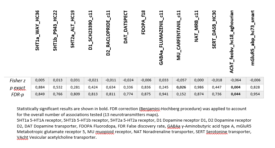
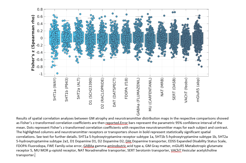
Results: This study included a cohort of 370 PD patients (mean age = 61.3 years, SD = 9.8; 65% male). 144 PD patients were classified as PD-pRBD. In the PD-pRBD group, voxel-based brain changes were found to be significantly associated with the spatial distribution of acetylcholine transporter (p=0.004; Table 1 and Figure 1), even after correcting for multiple comparisons (False Discovery Rate [FDR]-p=0.044). Furthermore, voxel-based brain changes in the PD-pRBD group, compared to the PD-noRBD group, were found to be significantly associated with the spatial distribution of mu-opioid receptors (p=0.026), but the effect did not survive correction for multiple comparisons (FDR-p=0.152).
Conclusion: Our study revealed an involvement of both the cholinergic and opioid systems in PD with RBD, through the indirect evaluation of neurotransmitter deficits. In addition, our findings highlight the potential of the JuSpace toolbox for assessing the underlying neurochemical alterations associated with nonmotor symptoms of Parkinson’s disease, using a radiation-free imaging modality.
References: 1. Jiménez-Jiménez, F.J., et al. Neurochemical Features of Rem Sleep Behaviour Disorder. Journal of Personalized Medicine, 2021. 11, DOI: 10.3390/jpm11090880.
2. Nomura, T., et al., Utility of the REM sleep behavior disorder screening questionnaire (RBDSQ) in Parkinson’s disease patients. Sleep Medicine, 2011. 12(7): p. 711-713.
3. Dukart, J., et al., JuSpace: A tool for spatial correlation analyses of magnetic resonance imaging data with nuclear imaging derived neurotransmitter maps. Human Brain Mapping, 2021. 42(3): p. 555-566.
To cite this abstract in AMA style:
D. Urso, B. Tafuri, S. Nigro, V. Gnoni, I. Rosenzweig, K. Ray Chaudhuri, G. Logroscino. Early neurotransmitters changes in Parkinson’s disease with REM sleep behaviour disorder [abstract]. Mov Disord. 2023; 38 (suppl 1). https://www.mdsabstracts.org/abstract/early-neurotransmitters-changes-in-parkinsons-disease-with-rem-sleep-behaviour-disorder/. Accessed December 25, 2025.« Back to 2023 International Congress
MDS Abstracts - https://www.mdsabstracts.org/abstract/early-neurotransmitters-changes-in-parkinsons-disease-with-rem-sleep-behaviour-disorder/


