Session Information
Date: Monday, June 20, 2016
Session Title: Surgical Therapy
Session Time: 12:30pm-2:00pm
Objective: To assess DTI connectivity patterns and correlations with weight gain in Parkinson’s disease patients after STN DBS.
Background: Weight gain is a well-known adverse effect of DBS, though the mechanism is poorly understood. We hypothesize that stimulation location and parameters affect different networks, impacting weight gain.
Methods: We examined weight change in a cohort of PD patients with monopolar STN DBS stimulation at 1, 2 and 3 mo post-operatively. We divided them into “significant gainers” (SG) and “non-gainers”(NG), based on a cut-off increase of 2 points in BMI. We modeled patient-specific DBS contacts using DBSproc1 and performed binary analysis to identify a connectivity profile for each group, using a 1% threshold of total connectivity (normalized by the total number of streamlines passing through the volumes of tissue activated). For patients whose active contacts remained the same for 1y, weighted average voltages and impedances were used to model an average connectivity profile. We correlated the rate of BMI change with connectivity measurements.
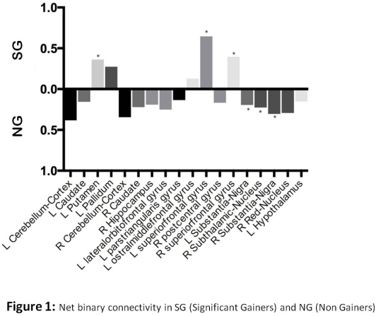
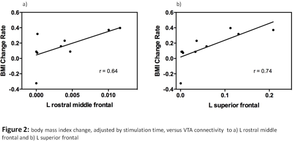
Results: 16 patients were included (11 SG, 5 NG). Mean BMI increased from 27.1 ±5 to 28.6 ±4.7 over 3 mo. Rapid weight gain occurred in the first 3 mo, with a plateau at 9-12 mo. SG had a significant increased connectivity to the L superior frontal cortex**, R superior frontal cortex*, L putamen*, and L rostral middle frontal cortex*. NG had a significant increased connectivity in the R STN**, and bilateral substantia nigra*. Active contacts remained unchanged in 10 subjects at 1y. In this subgroup, we found a correlation of BMI change with tracts to the L superior frontal and rostral middle frontal cortices. 

Conclusions: The pattern of weight gain after STN DBS in our patient population was characterized by a rapid initial weight gain followed by a plateau within the first year, consistent with previous observations. Using patient-specific VTA modeling in conjunction with DTI tractography, we identified brain regions related to BMI changes after bilateral STN DBS surgery. Our findings suggest that frontal and striatal modulations are related to weight changes. As a corollary, those who did not gain a substantial amount of weight seemed to have less relative extrastriatal connectivity. Further analysis is warranted, as this method could allow imaging-based prediction of weight gain and better understanding of its pathophysiologic mechanisms.
To cite this abstract in AMA style:
O.F. Ahmad, L. Huang, N. Vanegas-Arroyave, K. Zaghloul, S.G. Horovitz, C. Lungu. DTI tractographic correlates of weight gain in Parkinson’s disease patients after STN DBS [abstract]. Mov Disord. 2016; 31 (suppl 2). https://www.mdsabstracts.org/abstract/dti-tractographic-correlates-of-weight-gain-in-parkinsons-disease-patients-after-stn-dbs/. Accessed January 5, 2026.« Back to 2016 International Congress
MDS Abstracts - https://www.mdsabstracts.org/abstract/dti-tractographic-correlates-of-weight-gain-in-parkinsons-disease-patients-after-stn-dbs/
