Session Information
Date: Wednesday, September 25, 2019
Session Title: Neuroimaging
Session Time: 1:15pm-2:45pm
Location: Les Muses Terrace, Level 3
Objective: To evaluate the integrity of the nigro-striatal system with single photon emission computed tomography imaging with dopamine transporter (DaTscan) in patients with basal ganglia calcification (BGC) presenting with parkinsonism.
Background: BGC is reported in a variety of genetic, developmental, metabolic, infectious and other conditions. Parkinsonism is one of the most common movement disorders reported[1]; however, the underlying mechanism remains unclear. Studies of dopaminergic imaging in patients with BGC are scarce[2-3].
Method: We describe six patients with BGC followed in our Movement Disorders Unit. They all underwent detailed clinical examination and laboratory tests including phospho-calcium metabolism. Genetic analysis of SLC20A2, PDGFRB and PDGFB genes by next generation-sequencing was performed in those with family history. Brain calcification was evaluated with the total calcification score [4] and dopaminergic integrity was evaluated qualitatively with visual assessment.
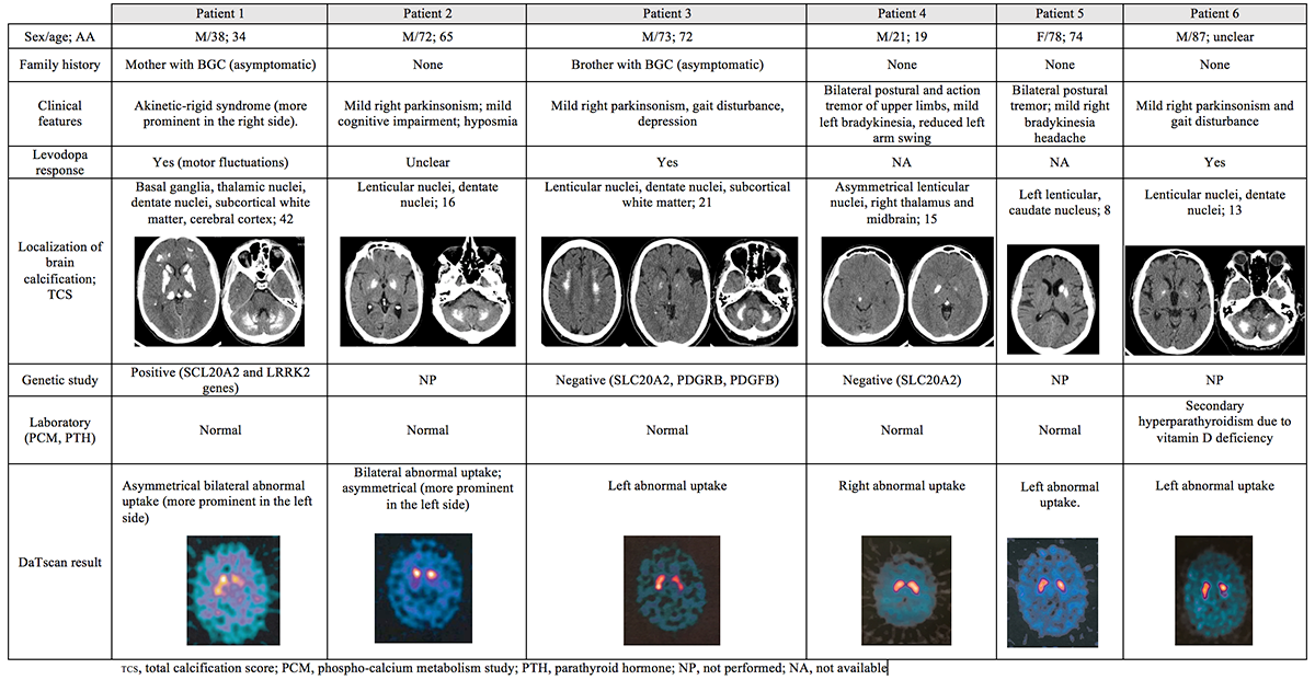
Results: Demographic, clinical and neuroimaging characteristics are summarized in [table1]. Four patients had an asymmetric parkinsonism and two a bilateral postural upper limb tremor and mild bradykinesia. Three had a documented effective response to levodopa and one an unclear response. All had bilateral BGC except one that had strictly unilateral calcification. Other localizations included: dentate nuclei, subcortical white matter, cortex and midbrain. We identified one patient with Primary Familial Brain Calcification due to an heterozygous mutation c.370G>A in the SLC20A2 gene and coincident mutation c.4321C Conclusion: We observed a correlation between positive DaTscans and clinical findings in our patients with BGC. We don’t know whether or not the calcification of the basal ganglia might affect the dopaminergic function or there is co-occurrence of different diseases. Further studies comparing DaTscan findings in symptomatic and asymptomatic patients with BGC are needed. References: [1] Manyam BV. What is and what is not “Fahr’s disease”. Parkinsonism Relat Disord 2005;11:73-80. [2] Caranci G, Frillea F, Barbato F, Brunetti A, Tassinari T, Di Ruzza M, Lena F, Gandolfi B, Castellano AE, Bartolo M, Colonnese C, Modugno N, Ruggieri S, Manfredi M. Scintigraphic, neuroradiological and clinical comparison in two patients with primary sporadic and two with secondary Fahr’s disease. Neurol Sci. 2011;32:337-341. [3] Koyama S, Sato H, Kobayashi R, Kwakatsu S, Kurimura M, Wada M, Kawanami T, Kato T. Clinical and radiological diversity in genetically confirmed primary familial brain calcification. Scientific Reports. 2017 [4] Nicolas G, Pottier C, Charbonnier C, et al. Phenotypic spectrum of probable and genetically-confirmed idiopathic basal ganglia calcification. Brain 2013; 136: 3395-3407. To cite this abstract in AMA style: « Back to 2019 International Congress MDS Abstracts - https://www.mdsabstracts.org/abstract/dopaminergic-imaging-in-patients-with-basal-ganglia-calcification-presenting-with-parkinsonism/

