Session Information
Date: Wednesday, September 25, 2019
Session Title: Neuroimaging
Session Time: 1:15pm-2:45pm
Location: Les Muses Terrace, Level 3
Objective: To investigate global resting-state functional connectivity (rsFC) patterns associated with response to L-DOPA and to subthalamic nucleus (STN) deep brain stimulation (DBS) in patients with advanced Parkinson’s disease (PD).
Background: Brain circuit dysfunction in PD involves an extensive global network [1–3]. A distinctive basal ganglia rsFC pattern has been linked with the ranked response to L-DOPA [4].
Method: Nineteen patients underwent 3-Tesla resting-state functional MRI (rsfMRI) in the ON-medication state prior to STN DBS. Improvement in UPDRS-III hemibody scores were assessed following L-DOPA therapy and STN DBS. Global FC was measured between regions-of-interest (ROIs) defined by the Automated Anatomical Labeling (AAL) atlas and the Montreal Neurologic Institute (MNI) PD25 subcortical atlas. Seed- and network-level correlations were made with an FDR-p<0.005. Tremor dominant (TD) and akinetic-rigid (AR) subgroups were also compared separately. Graph theoretical analysis was performed with an analysis threshold of FDR-p<0.005; and then looking at the top 15% of edges.
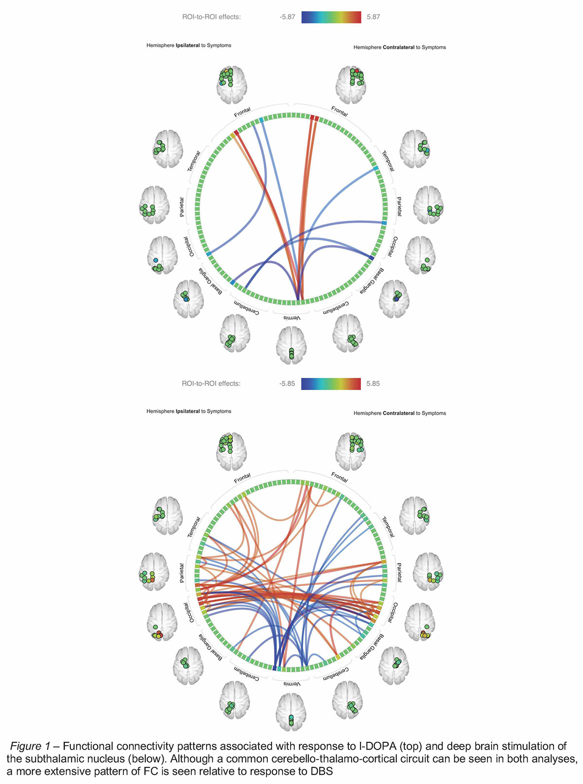
Results: Response to L-DOPA and to DBS displayed cerebellar desynchronization with bilateral thalami and synchronization with bilateral ventromedial prefrontal cortices (vmPFC). L-DOPA response was additionally associated with desynchronization between the vmPFC and the fusiform gyrus. Meanwhile, DBS response was associated with more widespread areas, which have been implicated in visuomotor control and planning [5–7][Fig. 1]. No significant differences in rsFC were seen between TD and AR groups. Graph theory analysis revealed that DBS response was inversely related to global efficiency of the thalamus and putamen bilaterally. No significant graph metrics were found relative to L-DOPA response.
Conclusion: Response to DBS and to L-DOPA share similar characteristics – particularly in cerebello-thalamo-cortical circuits, including those that play a role in planning, learning, decision-making, and reward-based behavior [8,9]. Preservation of distributed networks involved in visuomotor control and network integration of striatothalamocortical circuits appear to predict DBS response. These findings shed a light on the mechanism of action of DBS and L-DOPA and may help serve as useful treatment response biomarkers.
References: 1. Hou Y, Ou R, Yang J, Song W, Gong Q, Shang H: Patterns of striatal and cerebellar functional connectivity in early-stage drug-naïve patients with Parkinson’s disease subtypes. Neuroradiology 2018 Dec 22;60:1323–1333. 2. Suo X, Lei D, Li N, Cheng L, Chen F, Wang M, et al.: Functional Brain Connectome and Its Relation to Hoehn and Yahr Stage in Parkinson Disease. Radiology 2017 Dec;285:904–913. 3. Luo CY, Guo XY, Song W, Chen Q, Cao B, Yang J, et al.: Functional connectome assessed using graph theory in drug-naive Parkinson’s disease. J Neurol 2015 Jun 1;262:1557–1567. 4. Akram H, Wu C, Hyam J, Foltynie T, Limousin P, De Vita E, et al.: l-Dopa responsiveness is associated with distinctive connectivity patterns in advanced Parkinson’s disease. Mov Disord 2017;32. DOI: 10.1002/mds.27017 5. Göttlich M, Münte TF, Heldmann M, Kasten M, Hagenah J, Krämer UM: Altered resting state brain networks in Parkinson’s disease. PLoS One 2013 Oct 28;8:e77336. 6. Wu T, Ma Y, Zheng Z, Peng S, Wu X, Eidelberg D, et al.: Parkinson’s Disease—Related Spatial Covariance Pattern Identified with Resting-State Functional MRI. J Cereb Blood Flow Metab 2015 Nov 3;35:1764–1770. 7. Nackaerts E, Michely J, Heremans E, Swinnen S, Smits-Engelsman B, Vandenberghe W, et al.: Being on Target: Visual Information during Writing Affects Effective Connectivity in Parkinson’s Disease. Neuroscience 2018 Feb 10;371:484–494. 8. Caligiore D, Pezzulo G, Baldassarre G, Bostan AC, Strick PL, Doya K, et al.: Consensus Paper: Towards a Systems-Level View of Cerebellar Function: the Interplay Between Cerebellum, Basal Ganglia, and Cortex. Cerebellum 2017;16:203–229. 9. Reber J, Feinstein JS, O’Doherty JP, Liljeholm M, Adolphs R, Tranel D: Selective impairment of goal-directed decision-making following lesions to the human ventromedial prefrontal cortex. Brain 2017 Jun 1;140:1743–1756.
To cite this abstract in AMA style:
C. Wu, T. Foltynie, P. Limousin, L. Zrinzo, H. Akram. Distributed Global Functional Connectivity Networks Predict Responsiveness to L-DOPA and Subthalamic Deep Brain Stimulation [abstract]. Mov Disord. 2019; 34 (suppl 2). https://www.mdsabstracts.org/abstract/distributed-global-functional-connectivity-networks-predict-responsiveness-to-l-dopa-and-subthalamic-deep-brain-stimulation/. Accessed April 21, 2025.« Back to 2019 International Congress
MDS Abstracts - https://www.mdsabstracts.org/abstract/distributed-global-functional-connectivity-networks-predict-responsiveness-to-l-dopa-and-subthalamic-deep-brain-stimulation/

