Category: Parkinson's Disease: Neuroimaging
Objective: We used resting-state functional MRI (rs-fMRI) to investigate the alterations of functional connectivity (FC) of LC-related resting-state networks (RSNs) and the associations between RSNs changes and clinical symptoms in idiopathic rapid eye movement sleep behavior disorder (iRBD) and Parkinson’s Disease (PD) patients with (PDRBD+) and without RBD (PDRBD-).
Background: The locus coeruleus (LC) is severely affected in PD1. Most previous imaging studies have focused on the local degeneration of LC2, whereas the alterations of LC-related RSNs in the prodromal stage (iRBD) and different phenotypes of PD remain unclear.
Method: We recruited 53 iRBD, 58 PDRBD+ and 64 PDRBD- patients, and 69 healthy controls. We measured the between-group differences of FC between the LC and 5 RSNs, including networks of the default mode (DMN), executive control (ECN), salience (SAL), sensorimotor (SMN), and visual (VIS). We chose the pons as a reference region to calculate the FC between the pons and RSNs to validate that fMRI signals from surrounding regions did not significantly contaminate the FC results of LC-related RSNs. A partial correlation analysis was performed to explore the relationship between FC and clinical symptoms.
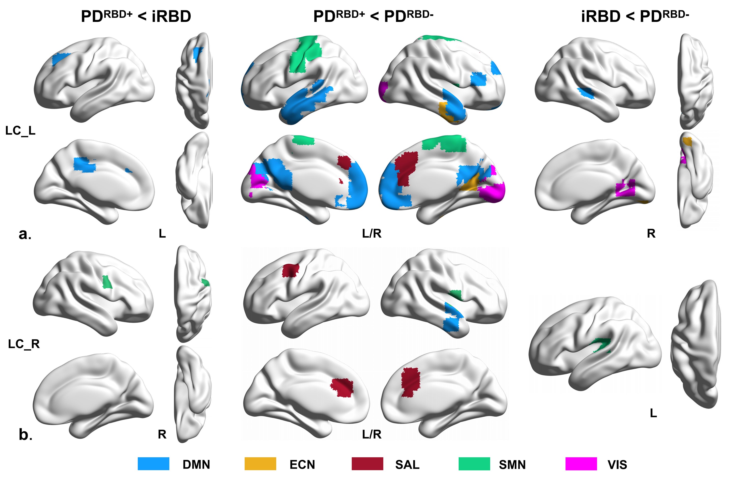
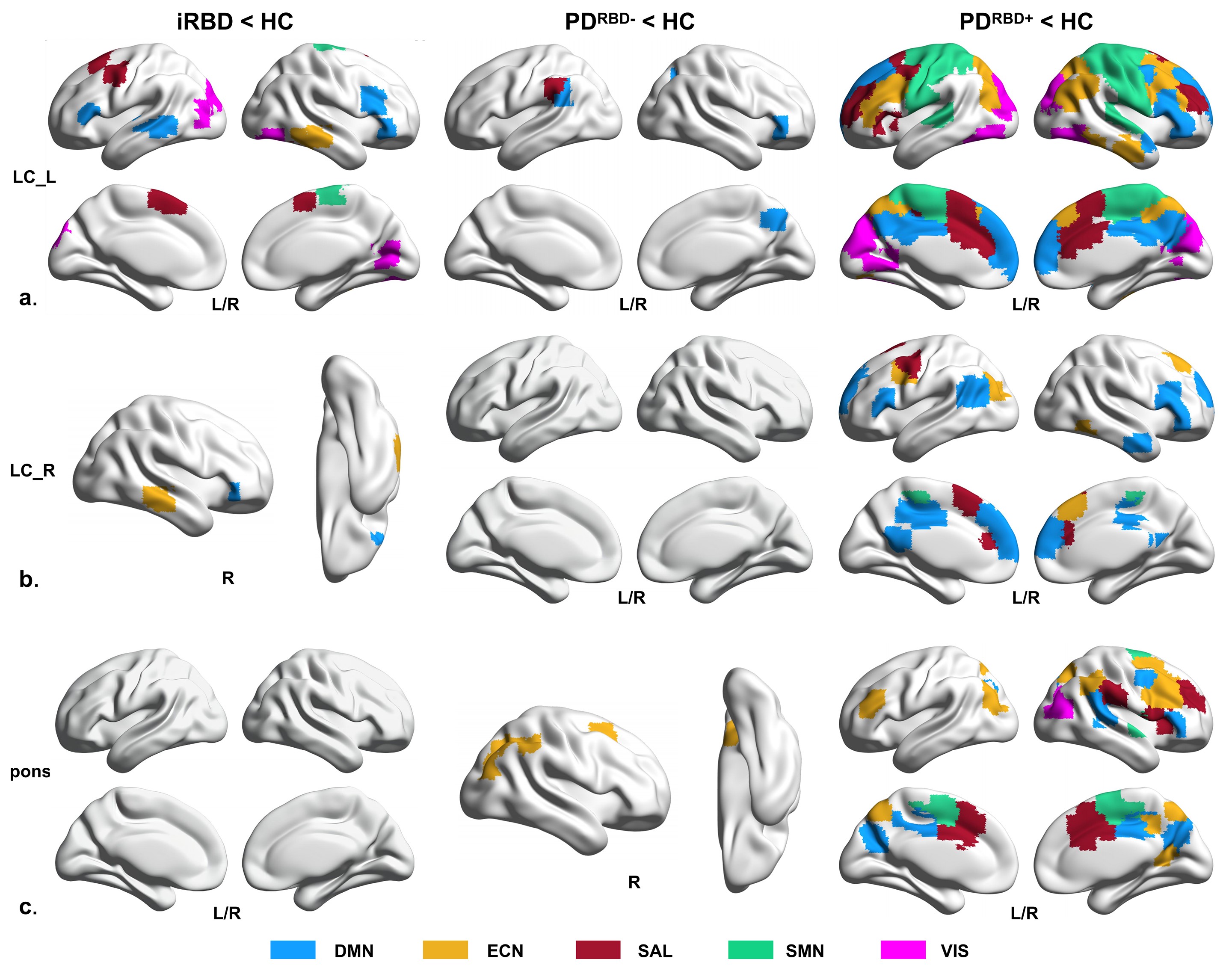
Results: The disrupted FC pattern of LC-related RSNs was similar in iRBD and PDRBD+ patients. PDRBD+ and iRBD patients exhibited more severe dysfunction of LC-related RSNs than PDRBD- patients (Fig. 1 and Fig. 2, FWE-corrected, cluster p < 0.05, cluster size > 9).
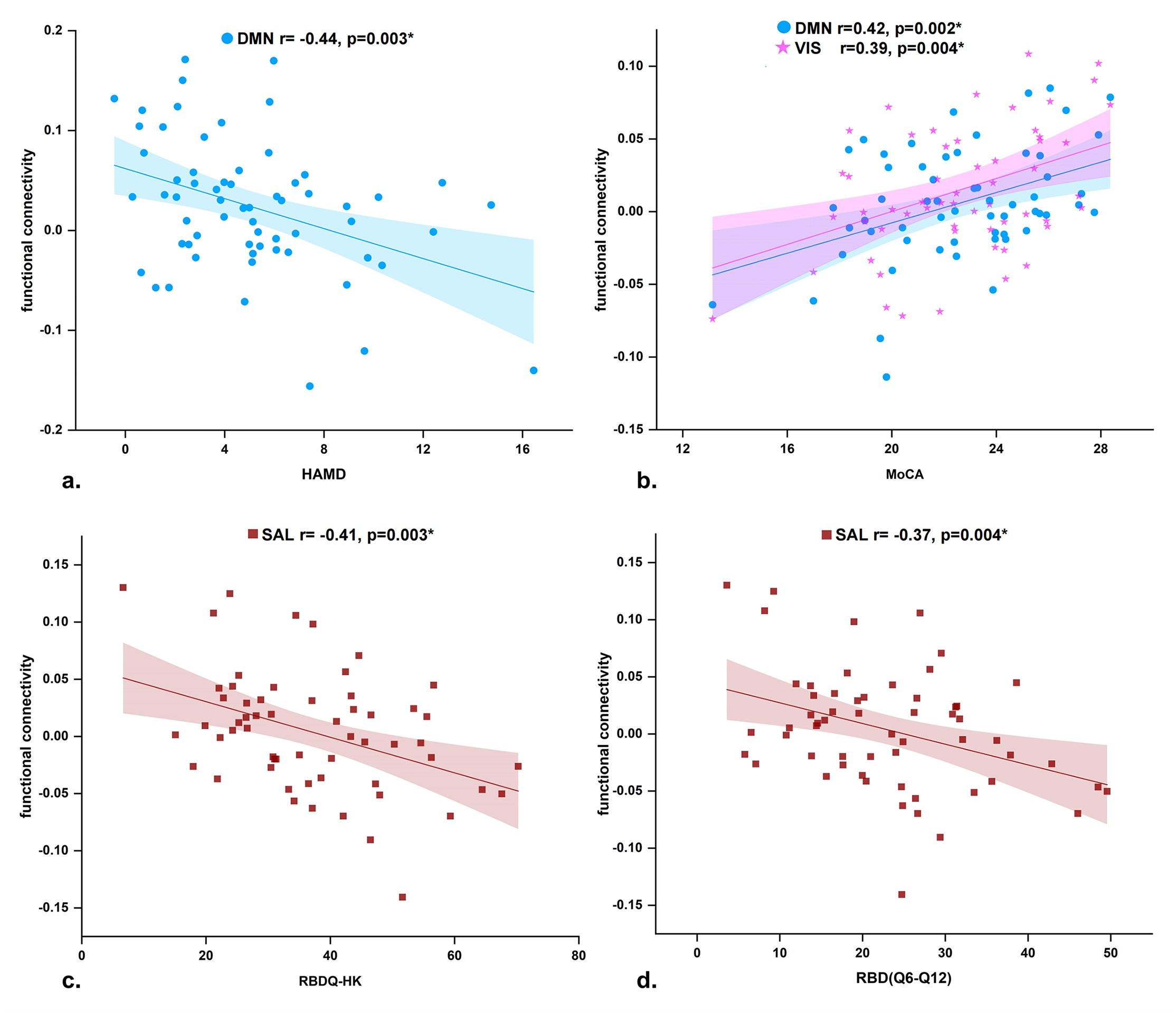
The FC of LC-related RSNs was correlated with cognition in iRBD and PDRBD+ patients, with disease duration in iRBD patients, with the severity of RBD in PDRBD+ patients, and with depression in PDRBD- patients (Fig. 3 and Fig. 4, p < 0.01, Bonferroni corrected).
Conclusion: The disrupted FC pattern of LC-related RSNs was similar in iRBD and PDRBD+ patients. PDRBD+ and iRBD patients exhibited more severe dysfunction of LC-related RSNs than PDRBD- patients (Fig. 1 and Fig. 2, FWE-corrected, cluster p < 0.05, cluster size > 9).
References: 1. Oertel, W. H., Henrich, M. T., Janzen, A. & Geibl, F. F. The locus coeruleus: Another vulnerability target in Parkinson’s disease. Mov. Disord. 34, 1423-1429 (2019).
2. Espay, A. J., LeWitt, P. A. & Kaufmann, H. Norepinephrine deficiency in Parkinson’s disease: the case for noradrenergic enhancement. Mov. Disord. 29, 1710-1719 (2014).
To cite this abstract in AMA style:
J. Sun, J. Ma, L. Gao, J. Wang, D. Zhang, L. Chen, J. Fang, T. Feng, T. Wu. Disruption of Locus Coeruleus-Related Functional Networks in Parkinson’s Disease [abstract]. Mov Disord. 2023; 38 (suppl 1). https://www.mdsabstracts.org/abstract/disruption-of-locus-coeruleus-related-functional-networks-in-parkinsons-disease/. Accessed February 8, 2026.« Back to 2023 International Congress
MDS Abstracts - https://www.mdsabstracts.org/abstract/disruption-of-locus-coeruleus-related-functional-networks-in-parkinsons-disease/


