Session Information
Date: Monday, October 8, 2018
Session Title: Parkinson's Disease: Neuroimaging And Neurophysiology
Session Time: 1:15pm-2:45pm
Location: Hall 3FG
Objective: test whether patients with Parkinson’s diseases (PD) and REM sleep behavior disorder (RBD) exhibit diminished state-related changes in their resting state fMRI global signal.
Background: Global resting state fMRI signals1 reflect state-dependent metabolic differences in brain networks, including external stimulus and vigilance changes between the eyes-closed and eyes-open states2.
Methods: 24 controls (CON, N=7 scanned twice), 51 PD patients (N=10 scanned twice), and 43 subjects with RBD(N=9 scanned twice) underwent MRI scanning (3T Philips) including a high resolution T1-weighted anatomical volume and separate eyes open and eyes closed resting state fMRI runs. Using AFNI, the fMRI volumes were motion corrected and aligned to the T1 volume. The mean resting state global signal was extracted for each run after motion-censoring (0.3 mm threshold) and regression of signal from ventricles and white matter. A single global signal value, defined as the standard deviation of the voxelwise signal across time averaged across all brain voxels, was computed for each eyes closed and eyes open run of each subject. The difference in open global signal was compared within each group of control, PD, and RBD subjects using a 2-tailed paired-test.
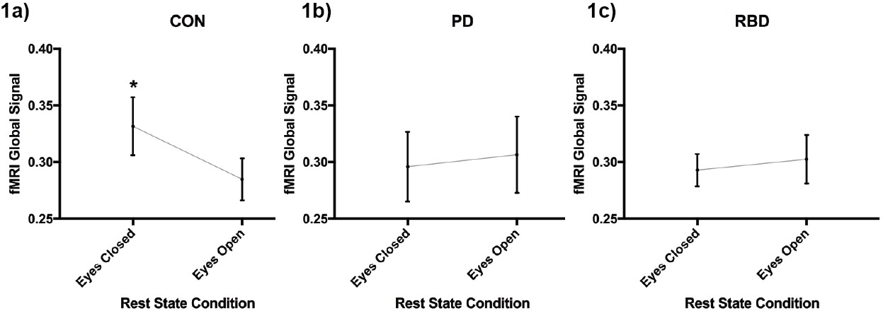
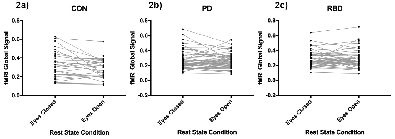
Results: The CON group showed a significant difference in the two resting state conditions with the global signal being higher during the eyes closed compared to eyes open condition (Fig 1a, t(30)=2.475, p=0.0192, 0.0469 mean of differences +/- 0.1055 SD). No significant differences were found between the eyes closed and eyes open conditions for the PD patients (Fig 1b, t(61)=0.2726, p=0.7861, 0.01061 mean of differences +/- 0.3039 SD) or subjects with RBD (Fig 1c, t(51)=0.529, p=5991, 0.00963 mean of differences +/- 0.1313 SD). Fig 2a-c show pattern of state changes for subjects in the CON, PD, and RBD groups respectively.
Conclusions: The difference obtained in controls is a replication consistent with patterns described in other studies showing increased global signal correlation amplitude in absence of external stimulation and during decreased vigilance. The average difference in global signal between eyes closed and eyes open conditions was diminished in both PD and RBD patients (no significant differences). Global resting state signals fail to respond to changes in external stimulation and/or changes in vigilance in these patients, which suggests that diminished state changes in the fMRI global signal in subjects at risk of developing a neurodegenerative disorder may be a biomarker both RBD and PD.
References: 1. Scholvinck ML, Maier A, Ye FQ, Duyn JH, Leopold DA. Neural basis of global resting-state fMRI activity. Proc Natl Acad Sci U S A. 2010;107(22):10238-10243. 2. Thompson GJ, Riedl V, Grimmer T, Drzezga A, Herman P, Hyder F. The Whole-Brain “Global” Signal from Resting State fMRI as a Potential Biomarker of Quantitative State Changes in Glucose Metabolism. Brain Connect. 2016;6(6):435-447.
To cite this abstract in AMA style:
J. Suescun, M. Schiess, L. Giancardo, R. Castriotta, T. Ellmore. Diminished fMRI global signal differences between eyes closed and eyes open resting state in RBD and PD [abstract]. Mov Disord. 2018; 33 (suppl 2). https://www.mdsabstracts.org/abstract/diminished-fmri-global-signal-differences-between-eyes-closed-and-eyes-open-resting-state-in-rbd-and-pd/. Accessed January 4, 2026.« Back to 2018 International Congress
MDS Abstracts - https://www.mdsabstracts.org/abstract/diminished-fmri-global-signal-differences-between-eyes-closed-and-eyes-open-resting-state-in-rbd-and-pd/


