Category: Parkinson's Disease: Neuroimaging
Objective: Patients with Parkinson’s disease (PD) experience changes in behavior, personality, and cognition. Mild Behavioral Impairment (MBI) is a validated neurobehavioral syndrome that identifies a high-risk group for incident cognitive decline by leveraging the risk associated with the emergence and persistence of neuropsychiatric symptoms in later life.
Background: The neurostructural underpinnings of MBI, as well as their differences with mild cognitive impairment (MCI), in PD remain poorly understood. Here we aimed to investigate the neurostructural correlates of MBI and MCI in PD using diffusion-weighted imaging.
Method: Participants included 91 PD patients and 36 healthy controls (HC). 69 PD patients did not have MBI (PD-nonMBI) and 22 patients had MBI (PD-MBI), whereas 53 patients had no MCI (PD-nonMCI) and 38 patients had MCI (PD-MCI). 10 PD patients had both MBI and MCI. We focused on connections from the hippocampus, amygdala, nucleus accumbens, caudate nucleus, and putamen to superior, middle, inferior, and orbito frontal gyri, and connectivity from the hippocampus and amygdala to other subcortical regions. Metrics of white matter integrity included free water (FW), free-water DTI-corrected tissue radial diffusivity (RDt), tissue fractional anisotropy (FAt) and fixel-based apparent fiber density (fixel-AFD). Diffusion-weighted and T1-weighted MRI data were analyzed using TractoFlow. All models for MBI were corrected for MCI status and vice versa.
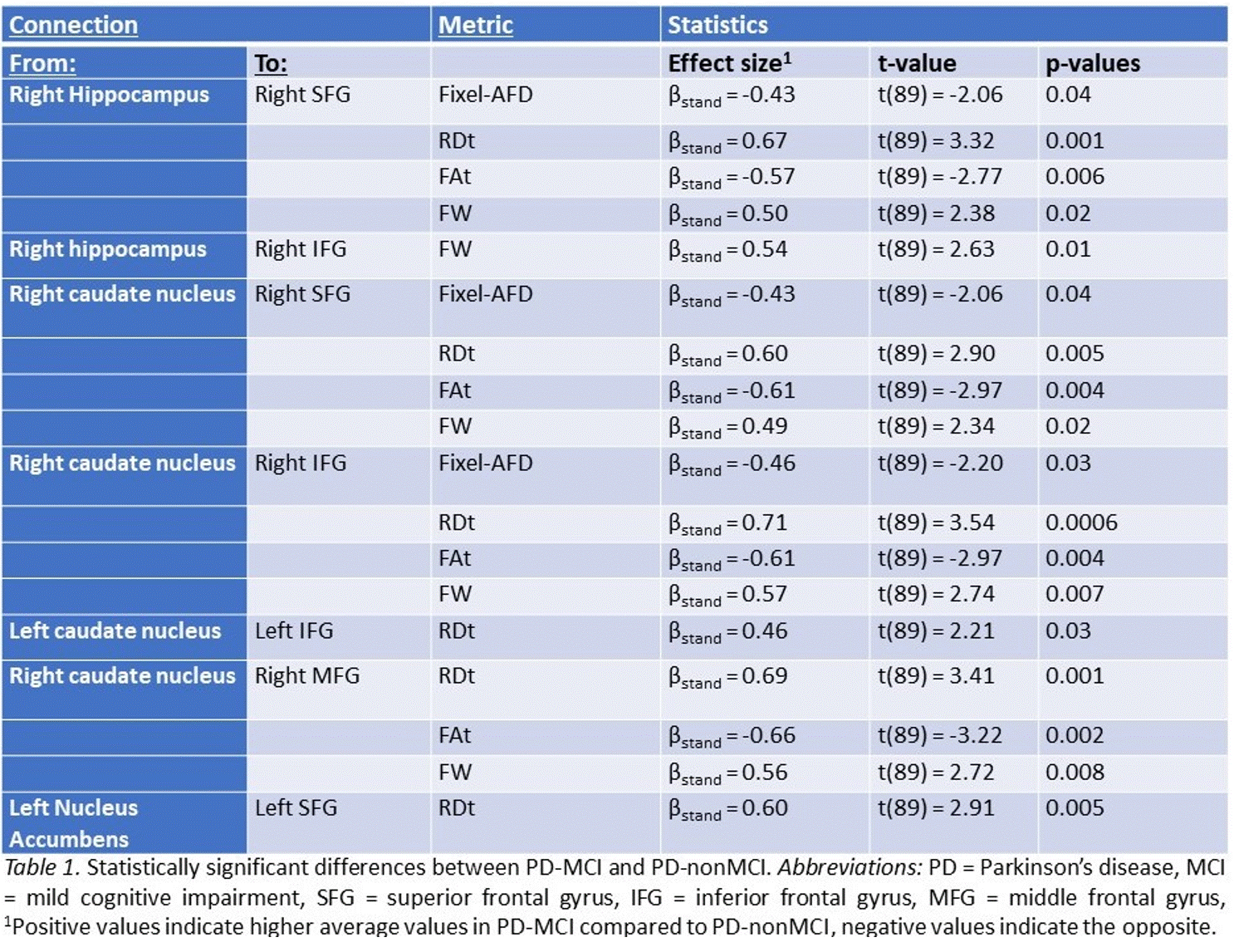
Results: Connections between left amygdala and putamen was disrupted in PD-MBI vs. PD-noMBI, as evidenced by reduced fixel-AFD, increased RDt, and reduced FAt. Impaired connectivity with the orbitofrontal gyrus (OFG) was found in PD-MBI vs. HC, as reflected by increased FW for the connections of (1) with left hippocampus, (2) with left nucleus accumbens, (3) with right amygdala, as well as increased RDt for connections between right OFG and amygdala. The microstructural correlates of PD-MCI involved decreased integrity of projections of the hippocampus and caudate nucleus to different frontal cortical regions (Table 1).
Conclusion: Connectivity between regions that are associated with neuropsychiatric symptoms including between amygdala and putamen, and between subcortical limbic regions and OFG, is disrupted in PD-MBI. PD-MCI, however, is related to impaired projections in regions associated with cognition between the caudate nucleus and hippocampus to multiple frontal regions.
To cite this abstract in AMA style:
H. Almgren, M. Descoteaux, M. Ghahremani, Z. Ismail, O. Monchi. Diffusion imaging study of mild behavioral impairment in Parkinson’s disease. [abstract]. Mov Disord. 2023; 38 (suppl 1). https://www.mdsabstracts.org/abstract/diffusion-imaging-study-of-mild-behavioral-impairment-in-parkinsons-disease/. Accessed February 4, 2026.« Back to 2023 International Congress
MDS Abstracts - https://www.mdsabstracts.org/abstract/diffusion-imaging-study-of-mild-behavioral-impairment-in-parkinsons-disease/

