Category: Ataxia
Objective: To report a case of acute anti-Yo positive paraneoplastic cerebellar degeneration (PCD) with atypical brain MRI findings.
Background: PCD is a rare disease but accounted for the highest prevalence among paraneoplastic neurological syndromes. Anti-Yo antibody is the most commonly detected onconeural antibody in patients with PCD and is usually associated with breast and gynecological malignancies. Based on clinical features, it is difficult to differentiate acute PCD from other acute cerebellar syndromes such as virus associated cerebellitis. Brain MRI in anti-Yo positive PCD is often normal in the early stages and cerebellar atrophy can be seen several months after the onset of symptoms.
Method: A case report
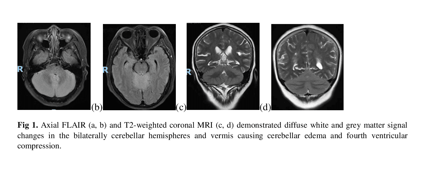
Results: A 55-year-old female patient presented to our department with one-week history of acute onset of vertigo, vomiting, slurred speech, and gait disorder. Her medical history was unremarkable. Neurological examination showed nystagmus, scanning dysarthria, limb dysmetria, truncal ataxia, head titubation, bilateral extensor plantar responses and normal tendon reflexes. Brain MRI performed at seventh day of the onset of symptoms revealed diffuse edema of bilateral cerebellar hemispheres and vermis with T2W hyperintensity and T1W hypointensity without gadolinium enhancement (figure 1). Cerebrospinal fluid (CSF) analysis showed pleocytosis (42 cells/mm3 and lymphocytes 95%), elevated protein level (71.43 mg/dl) and increased lactate level (3.93 mmol/l). Gram stain and CSF culture did not show any bacteria, fungi or parasites. Her CSF virology polymerase chain reaction results were negative. A complete blood count, electrolytes, renal, hepatic, thyroid function and B12 level tests were normal. Blood tests for tumor markers showed elevated carbohydrate antigens (CA125 = 353.9 U/mL, CA 15-3 = 33.9 U/mL, and CA 19-9 = 57.1 U/mL). Immunologic tests for HIV types 1 and 2, ANA, RPR, anti-GAD, anti-MMDA receptor, and lupus anticoagulant were all negative. Anti-neuronal antibodies in the serum were positive for anti-Yo antibodies. CT of the pelvis demonstrated a right ovarian tumor and paraaortic lymph nodes.
Conclusion: Our clinical case revealed that diffuse cerebellar edema on MRI can be seen in the early stage of PCD associated anti-Yo and acute onset cerebellar ataxia patients with uncertain cause should be screened for onconeural antibodies.
References: [1]..Giometto B, Grisold W, Vitaliani R, et al.(2010).Paraneoplastic neurologic syndrome in the PNS Euronetwork database: a European study from 20 centers. Arch Neurol. 67(3), 330-5;
[2].Shams’ili S, Grefkens J, de Leeuw B, et al.(2003).Paraneoplastic cerebellar degeneration associated with antineuronal antibodies: analysis of 50 patients. Brain. 126(Pt 6), 1409-18;
[3]..Venkatraman A and Opal P(2016).Paraneoplastic cerebellar degeneration with anti-Yo antibodies – a review. Ann Clin Transl Neurol. 3(8), 655-63;
To cite this abstract in AMA style:
T. Dang, K. Vo, U. Ha, Q. Pham, C. Phan, T. Tran. Diffuse cerebellar edema in acute anti-Yo positive paraneoplastic cerebellar degeneration: A case report [abstract]. Mov Disord. 2023; 38 (suppl 1). https://www.mdsabstracts.org/abstract/diffuse-cerebellar-edema-in-acute-anti-yo-positive-paraneoplastic-cerebellar-degeneration-a-case-report/. Accessed February 6, 2026.« Back to 2023 International Congress
MDS Abstracts - https://www.mdsabstracts.org/abstract/diffuse-cerebellar-edema-in-acute-anti-yo-positive-paraneoplastic-cerebellar-degeneration-a-case-report/

