Category: Dystonia: Pathophysiology, Imaging
Objective: To find electrophysiological biomarkers which differ cervical dystonia patients with jerky and sinusoidal head tremor.
Background: Two common movement disorders, dystonia and tremor, frequently overlap [1]. Their biological relationships remain unclear. One hypothesis is that tremor and dystonia are due to the same fundamental biological process. A second hypothesis is that tremor and dystonia are due to distinct biological processes which frequently co-occur. Here we test these hypotheses by measuring physiological activity of the globus pallidus in the most common form of dystonia, cervical dystonia, which frequently combines with tremor of the head.
Method: We compared three groups of patients, those with no tremor at all (pure dystonia), those with jerky head oscillations (dystonic tremor), and those with sinusoidal tremor. We used microelectrode recording from 13 CD subjects undergoing deep brain stimulation (DBS) surgery under local anesthesia. We analyzed the spontaneous activity of 727 pallidal neurons. Clustering of spike-train recordings allowed classification of pallidal activity according to burst, pause, and tonic neurons – the types of neurons expected in dystonia patients.
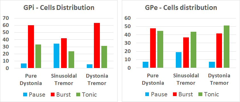
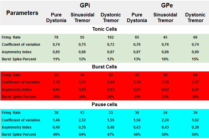
Results: We found the differences in cells distribution between analyzed groups of patients [figure1]. Burst GPi neurons were more common in pure dystonia or dystonic tremor groups compared to the sinusoidal tremor group. Pause GPi and GPe neurons were more common in the sinusoidal tremor group compared to the pure dystonia or dystonic tremor groups. In addition to their frequent prevalence, the pause neurons in sinusoidal tremor group had lower firing rate and were more irregular. The number of spikes within the burst were higher and inter-burst intervals were longer in sinusoidal tremor group. Neurons of patients with pure dystonia and dystonic tremor had similar parameters [table1]. We also found bihemispheric asymmetry in the dystonic tremor and pure dystonia groups, while such asymmetry was not present in the sinusoidal oscillation group.
Conclusion: In summary, our results suggest biological differences in the basal ganglia network behavior in pure cervical dystonia or cervical dystonia combined with irregular head oscillations compared to cervical dystonia with sinusoidal oscillations.
These data were presented at SFN-2019. The study was funded by the Russian Science Foundation (project 18-15-00009).
References: 1. Shaikh AG, Zee DS, Jinnah HA. Oscillatory head movements in cervical dystonia: Dystonia, tremor, or both? Mov Disord. 2015 May;30(6):834-42
To cite this abstract in AMA style:
A. Sedov, S. Usova, U. Semenova, A. Gamaleya, A. Tomskiy, H. Jinnah, A. Shaikh. Difference in pallidal single unit activity in cervical dystonia with jerky and sinusoidal head tremor [abstract]. Mov Disord. 2020; 35 (suppl 1). https://www.mdsabstracts.org/abstract/difference-in-pallidal-single-unit-activity-in-cervical-dystonia-with-jerky-and-sinusoidal-head-tremor/. Accessed January 4, 2026.« Back to MDS Virtual Congress 2020
MDS Abstracts - https://www.mdsabstracts.org/abstract/difference-in-pallidal-single-unit-activity-in-cervical-dystonia-with-jerky-and-sinusoidal-head-tremor/


