Category: Parkinson's Disease: Neurophysiology
Objective: To detect the presence of Levodopa induced Dyskinesia (LID) in Parkinson’s disease (PD) patients based on spectral characteristics from invasive cortico-subthalamic recordings using machine learning.
Background: Adaptive DBS (aDBS) in Parkinson’s disease (PD) automatically tailors DBS-parameters according to patients’ real-time symptom states. While beta-frequency related biomarkers are widely reproduced as biomarkers for the medication-OFF state [1], biomarkers describing the medication-ON state gained more attention only recently [2, 3]. Gamma-frequency features are linked to LID [2] and especially electrocorticography (ECoG) holds potential to capture this biomarker. Here, we aim to find an aDBS biomarker for the ON-medication state by detecting LID-presence based on STN LFP and ECoG recordings in PD patients using machine learning modeling.
Method: Six PD patients with externalized bilateral STN DBS electrodes and a unilateral ECoG-strip were included. The ECoG-strip covered parts of the sensorimotor cortex.
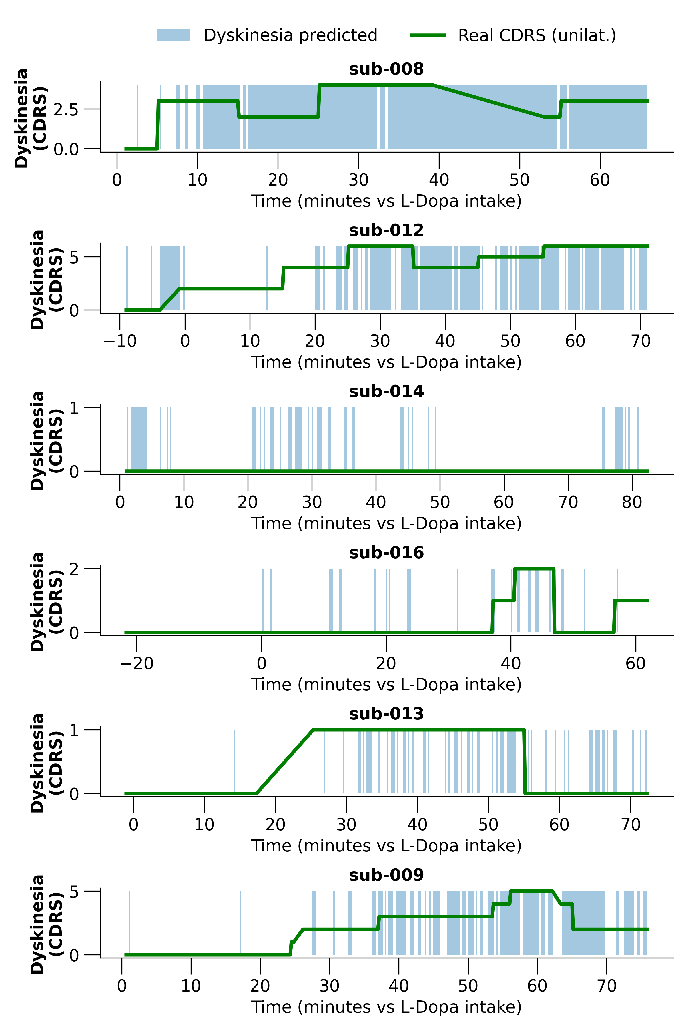
During this protocol, subjects started in a standardised OFF-state and received +/- 200 mg fast-acting levodopa. Over an average time course of one hour, the subjects performed repetitively rest and self-paced hand-tapping tasks. 5 out of 6 subjects developed LID contralateral to the ECoG-strip. We excluded moments with LID only ipsilateral to the ECoG-strip here.
We included unilateral, multichannel STN LFP- and ECoG-signals and filtered both separately using alpha, beta, and gamma specific spatial-spectral-decompostions. For every 10-second window, the maximum- and mean-peak values of the frequency-specific spectral peaks, and the coefficient of variation of the filtered time series were included as features.
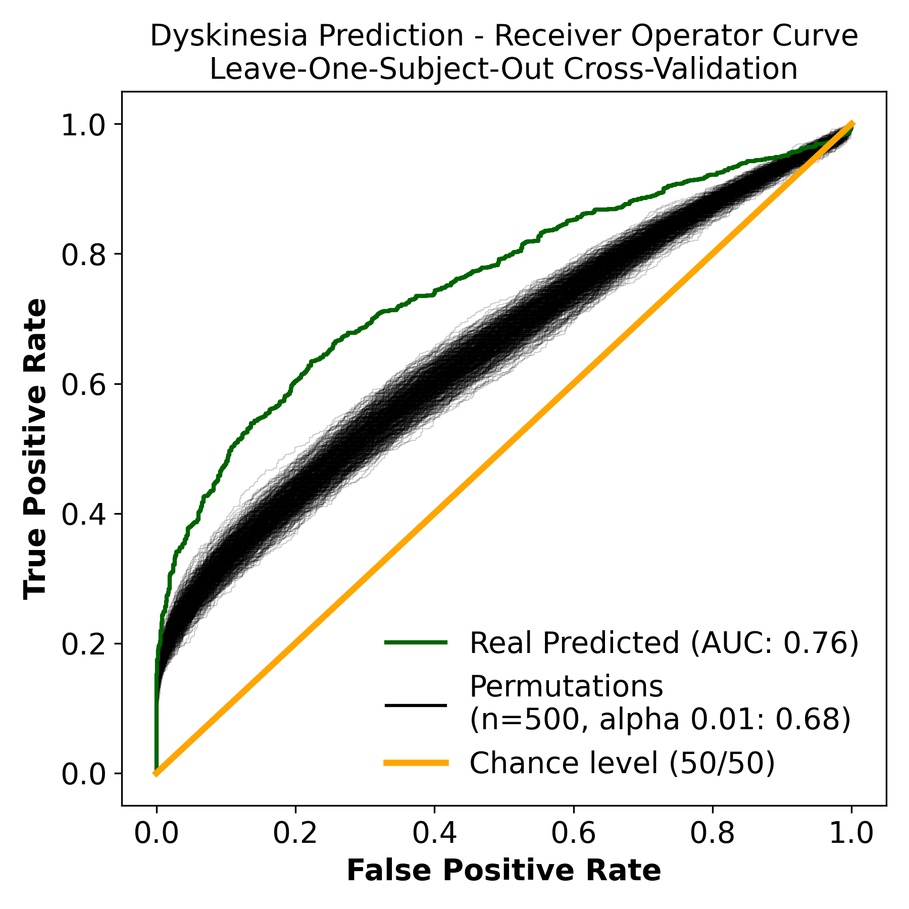
Predictive modeling was done with a logistic regression model in a Leave-One-Subject-Out cross-validation, and a permutation-test (n=500) was done for significance testing.
Results: LID-presence was predicted with an area under the curve of 0.76 (p < 0.01) and an accuracy of 0.70 [Figure 1]. The performance was good for both patients with severe as well as no LID [Figure 2].
Conclusion: LID could be detected significantly based on short-windowed invasive cortico-subthalamic recordings. These results can lead to aDBS-biomarkers representing the medication-ON state and thereby improving the usability and personification of aDBS.
References: [1]: van Wijk et al. A systematic review of local field potential physiomarkers in Parkinson’s disease: from clinical correlations to adaptive deep brain stimulation algorithms. J Neurol. 2023 Feb;270(2):1162-1177.
[2]: Wiest et al. Finely-tuned gamma oscillations: Spectral characteristics and links to dyskinesia. Exp Neurol. 2022 May;351:113999.
[3]: Gilron et al. Long-term wireless streaming of neural recordings for circuit discovery and adaptive stimulation in individuals with Parkinson’s disease. Nat Biotechnol. 2021 Sep;39(9):1078-1085.
To cite this abstract in AMA style:
J. Habets, R. Lofredi, J. Busch, V. Mathiopoulou, J. Roediger, R. Koehler, J. Vanhoecke, P. Krause, K. Faust, GH. Schneider, WJ. Neumann, A. Kühn. Detection of Levodopa induced Dyskinesia based on spectral features from subthalamic-LFP and sensorimotor-ECoG [abstract]. Mov Disord. 2023; 38 (suppl 1). https://www.mdsabstracts.org/abstract/detection-of-levodopa-induced-dyskinesia-based-on-spectral-features-from-subthalamic-lfp-and-sensorimotor-ecog/. Accessed April 20, 2025.« Back to 2023 International Congress
MDS Abstracts - https://www.mdsabstracts.org/abstract/detection-of-levodopa-induced-dyskinesia-based-on-spectral-features-from-subthalamic-lfp-and-sensorimotor-ecog/


