Session Information
Date: Thursday, June 8, 2017
Session Title: Parkinson's Disease: Neuroimaging And Neurophysiology
Session Time: 1:15pm-2:45pm
Location: Exhibit Hall C
Objective:
To investigate pooled effect of STN-DBS effect on brain activity in Parkinson’s disease (PD), and to address the result inconsistency across previous functional image studies.
Background: Deep brain stimulation of subthalamic nucleus (STN-DBS) has been becoming an effective treatment strategy for patients with PD. However, the biological mechanism underlying the DBS treatment remains poorly understood. Functional brain imaging techniques provide unique opportunities to ascertain how STN-DBS modulates brain activity, but results often varied across studies. Activation likelihood estimation(ALE) meta-analysis allows a quantitative summarization by synthesizing a spatial activation map based on reported coordinates.
Methods: In the present study we performed ALE meta-analysis in which functional imaging studies concerning STN-DBS effects on either resting-state or task-evoked brain activity in PD were searched. The STN-DBS meta-analyses were conducted by using GingerALE version 2.3.3 (http://brainmap.org/ale/). We further performed functional connectivity analysis in healthy individuals. To our knowledge, this is the first ALE meta-analysis that explored STN-DBS effect on brain activity.
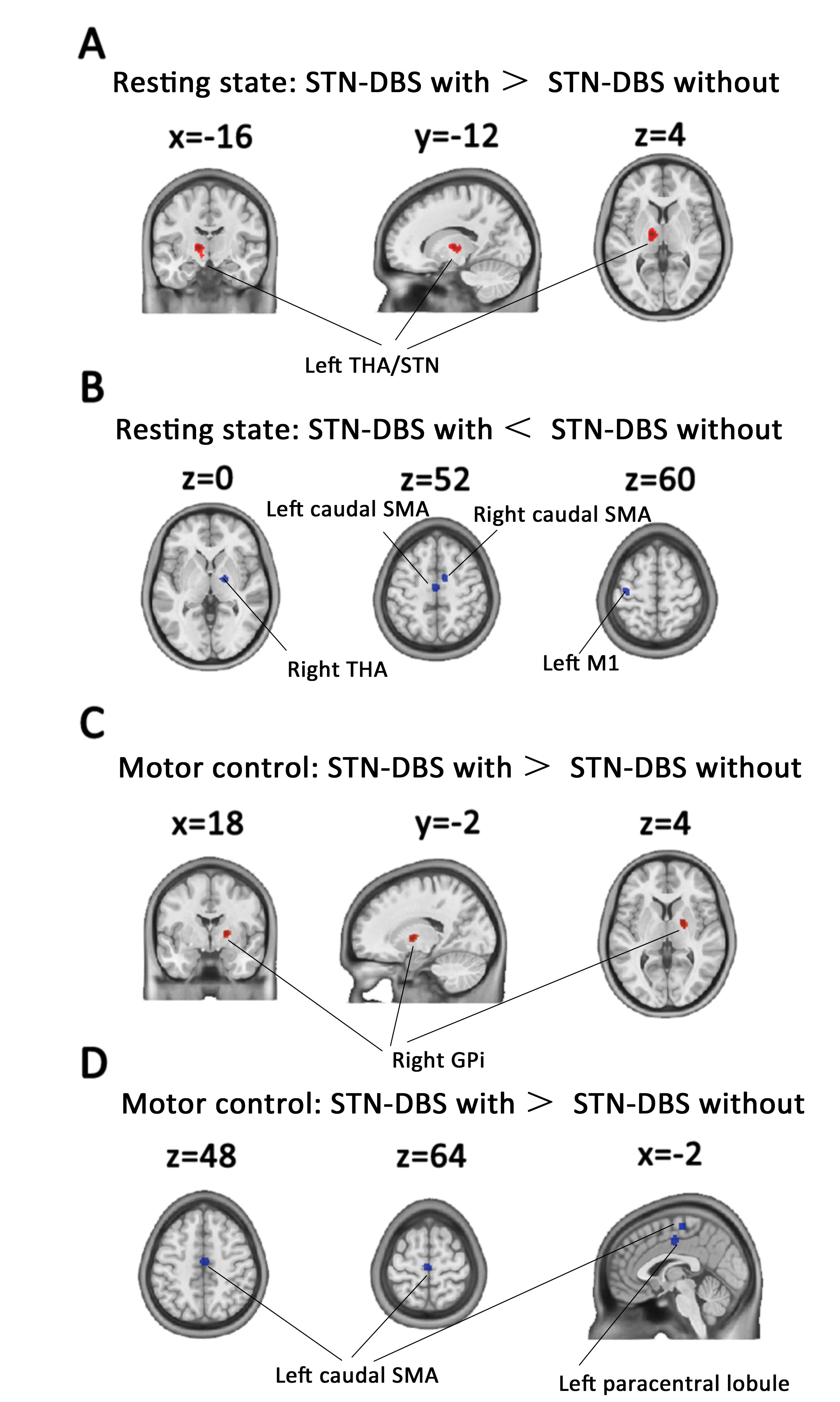
Results: First, meta-analysis based on thirteen resting-state functional imaging studies in Parkinson’s disease revealed that STN-DBS elevated brain activity in the STN and thalamus, and reduced activity in the caudal supplementary area and primary motor cortex during rest (Figure 1). Functional connectivity analysis based on a group of resting-state functional MRI data in healthy adults revealed that these treatment-related brain areas were functionally connected within a motor-related circuit. Second, meta-analysis based on five motor-control functional imaging studies in Parkinson’s disease revealed that STN-DBS elevated brain activities in the globus pallidus internus, and decreased activities in the caudal supplementary area and paracentral lobule (Figure 1).
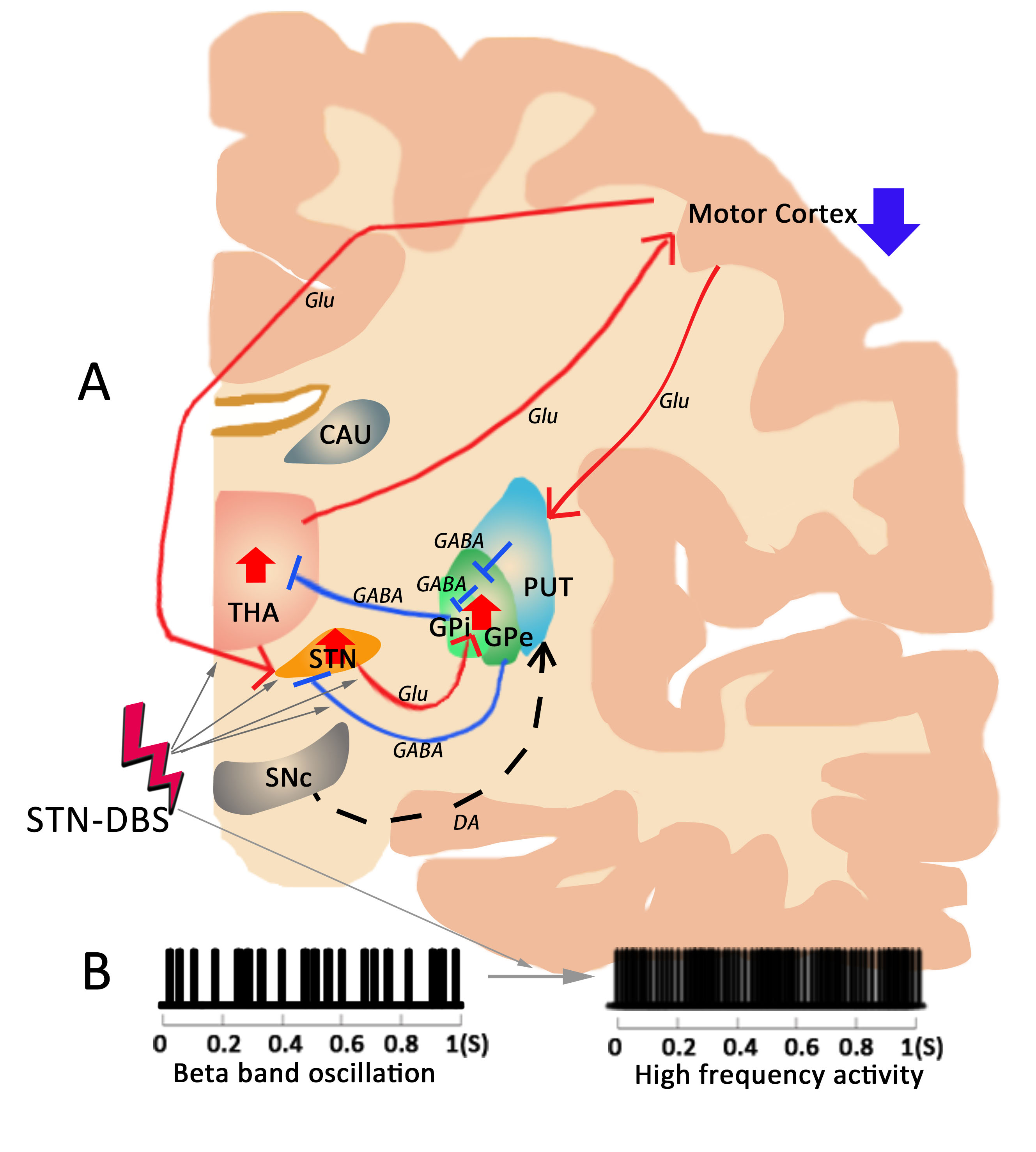
Conclusions: STN-DBS increased brain activity in the subcortical regions (i.e. STN, GPi and thalamus), and decreased brain activity in the motor cortex (i.e. SMA and M1). In conclusion, we postulate that STN-DBS may activate the basal ganglia and disrupt the information flow in the STC circuit in the meanwhile (Figure 2).
To cite this abstract in AMA style:
H. Chen. Deep Brain Stimulation of Subthalamic Nucleus Selectively Modulates Striato-Thalamo-Cortical Circuit in Parkinson’s Disease: a Systematic Review and ALE Meta-analysis [abstract]. Mov Disord. 2017; 32 (suppl 2). https://www.mdsabstracts.org/abstract/deep-brain-stimulation-of-subthalamic-nucleus-selectively-modulates-striato-thalamo-cortical-circuit-in-parkinsons-disease-a-systematic-review-and-ale-meta-analysis/. Accessed February 6, 2026.« Back to 2017 International Congress
MDS Abstracts - https://www.mdsabstracts.org/abstract/deep-brain-stimulation-of-subthalamic-nucleus-selectively-modulates-striato-thalamo-cortical-circuit-in-parkinsons-disease-a-systematic-review-and-ale-meta-analysis/


