Session Information
Date: Wednesday, September 25, 2019
Session Title: Neuroimaging
Session Time: 1:15pm-2:45pm
Location: Les Muses Terrace, Level 3
Objective: To study any inconsistency in the neural connections responsible for facilitating spatial recognition of olfactory stimuli as observed in people diagnosed with Parkinson’s Disease (PD) compared to those not affected by PD, and during the progression of the same.
Background: Olfactory dysfunctions are observed in approximately 90% of people affected by PD. Like in most neurodegenerative disorders, olfactory dysfunction is a clinical marker appearing years before the declining motor and cognitive functions in PD. The temporal-parietal junction (TPJ) contributes to spatial recognition, and lesions in this region or disruption of its neural communication have been associated with lack of spatial attention to various stimuli including visual, auditory, and less popularly olfactory.
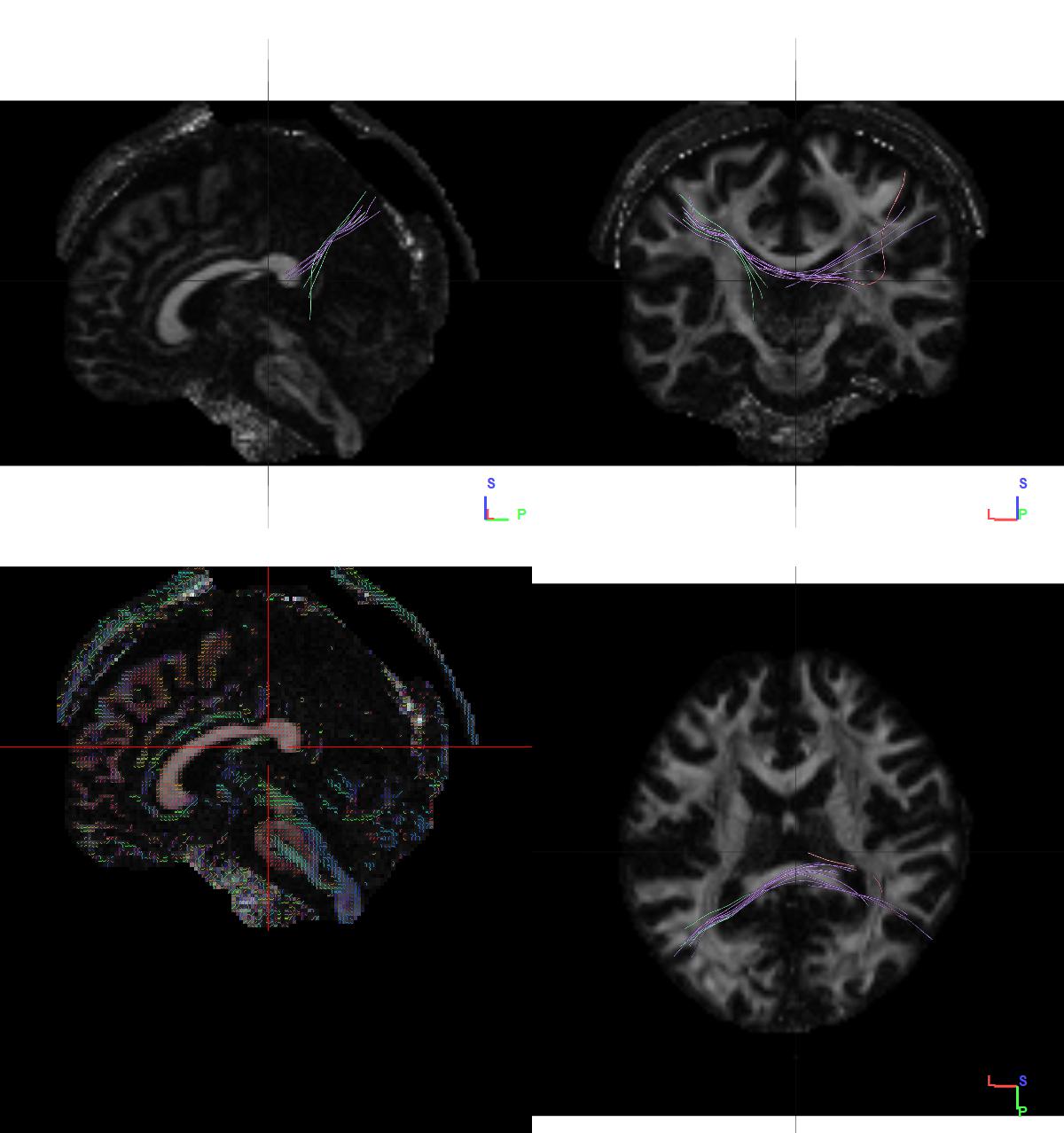
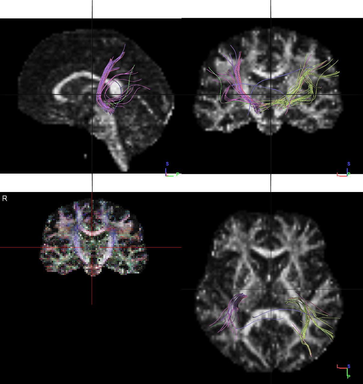
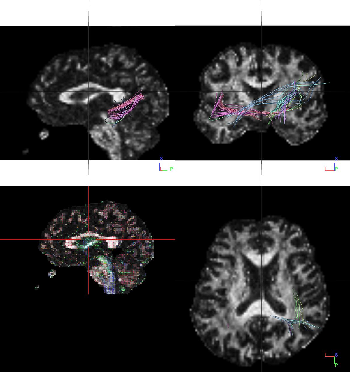
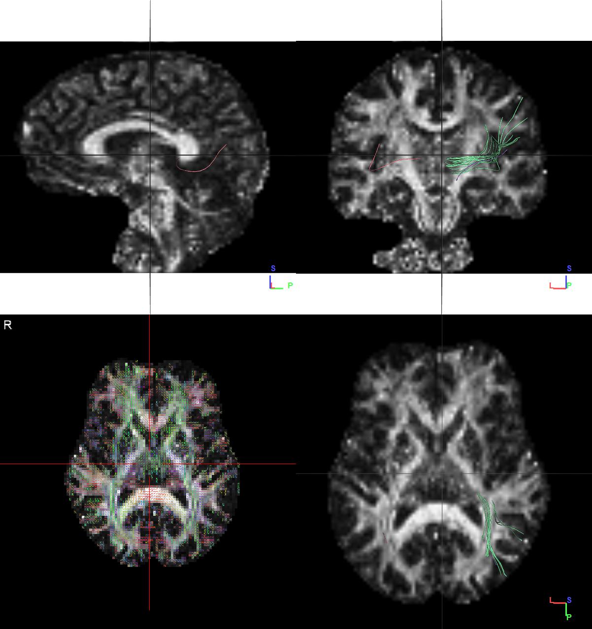
Method: This study applies fibre tractography using Diffusion Tensor Imaging. The neural connection between the olfactory cortex and TPJ have been studied in Magnetic Resonance Imaging (MRI) scans from four different groups of people including a control group, and three groups of people diagnosed with PD from widely spaced stages of progression of the disease. The study has been conducted with MRI scans obtained from 40 different people for each group.
Results: A significant difference in the number of fibres connecting the two regions was noted between the control group and the groups affected with PD (figure 1-4). A p-value of 0.037 was obtained. As the stages progressed, a decrease by 20 fibres on an average was noted and a p-value of 0.0001 was obtained.
Conclusion: Upholding the hypothesis, a significant difference in the number of fibres have been seen between the control and PD affected groups, and the further decline has been observed in the progressive stages of PD. This suggests that monitoring the neural connectivity between the olfactory cortex and TPJ or monitoring the neural connections facilitating one’s spatial attention to olfactory stimuli can provide for a way to monitor the progression of PD or a mode of staging on the basis of olfactory dysfunction progression.
References: [1] Doty RL. Olfactory dysfunction in Parkinson disease. Nat Rev Neurol. 2012 May 15;8(6):329-39. [2] Lamb MR., et al. Attention and interference in the processing of global and local information: Effects of unilateral temporal-parietal junction lesions. Neuropsychologia 27(4). 471-483. [3] Scholz J., et al. Distinct Regions of Right Temporo-Parietal Junction Are Selective for Theory of Mind and Exogenous Attention. PLoS One 4(3), e4869. Pierre and Marie Curie University. [4]Friedrich FJ., et al. Spatial attention deficits in humans: a comparison of superior parietal and temporal-parietal junction lesions. Neuropsychology 12(2). 193-207.
To cite this abstract in AMA style:
H. Chatterjee, G. Elumalai, N. Osakwe, N. Sewram. Decline in Neural Connectivity in Olfactory Spatial Attention: A Comparative Study in the Progression of Parkinson’s Disease [abstract]. Mov Disord. 2019; 34 (suppl 2). https://www.mdsabstracts.org/abstract/decline-in-neural-connectivity-in-olfactory-spatial-attention-a-comparative-study-in-the-progression-of-parkinsons-disease/. Accessed April 25, 2025.« Back to 2019 International Congress
MDS Abstracts - https://www.mdsabstracts.org/abstract/decline-in-neural-connectivity-in-olfactory-spatial-attention-a-comparative-study-in-the-progression-of-parkinsons-disease/


