Session Information
Date: Tuesday, September 24, 2019
Session Title: Parkinsonisms and Parkinson-Plus
Session Time: 1:45pm-3:15pm
Location: Agora 3 West, Level 3
Objective: The present study aimed at establishing clinical and brain functional patterns at FDG-PET that could distinguish Corticobasal Syndrome(CBS) variants due to AD from Tau pathology based on Pittsburgh Compound B positron emission tomography(PiB-PET) status.
Background: CBS is an atypical parkinsonian syndrome first considered a motor disorder but now also recognized as a cognitive disorder. The term CBS denotes the phenotype of multiple pathologies, including Alzheimer’s disease (AD) and 4-repeat Tauopathies. Accurate antemortem diagnosis is challenging and diagnostic methods are being developed to predict the underlying pathology.
Method: Twenty-one patients meeting criteria for probable CBS[1] were evaluated concerning their movement disorders and cognitive symptoms. They were submitted to FDG-PET and, according to its patterns, distributed into an AD group (likely related to AD) and a non-AD group (likely unrelated to AD). A small subset of 10 patients underwent PiB-PET and was compared by positive or negative status. Clinical profile and FDG scans were analyzed blinded to PiB-PET results.
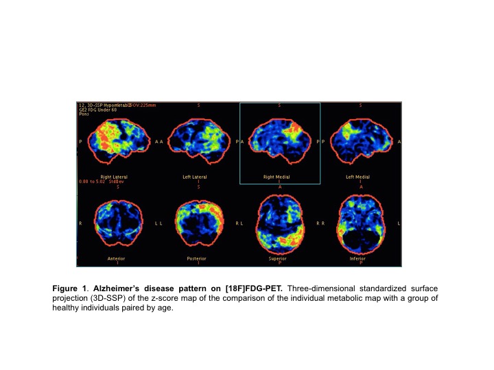
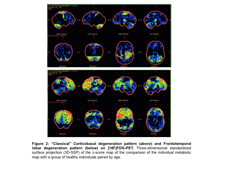
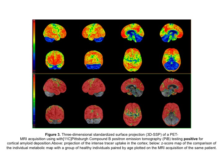
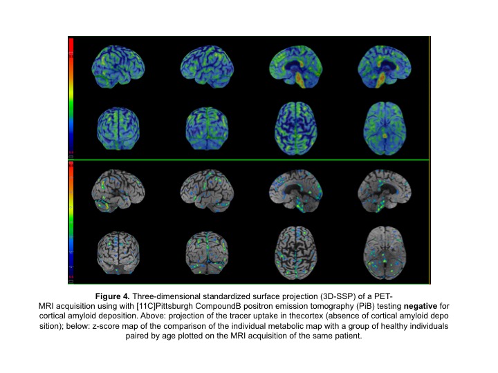
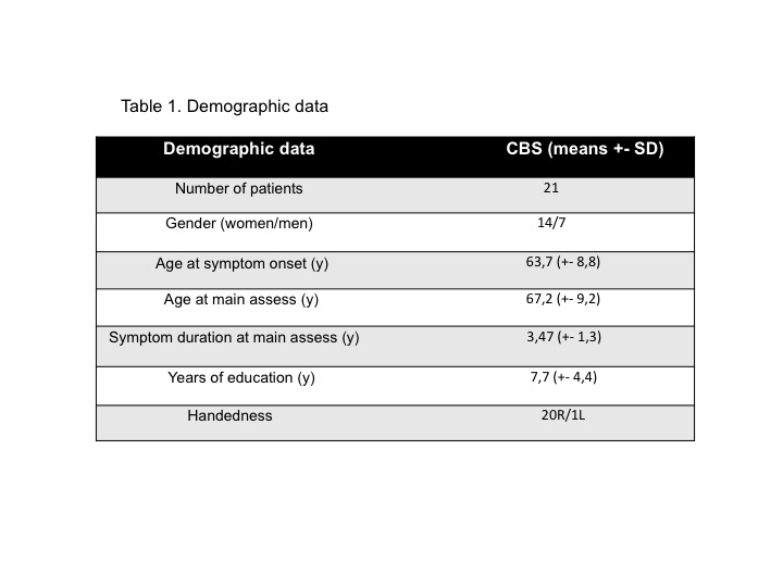
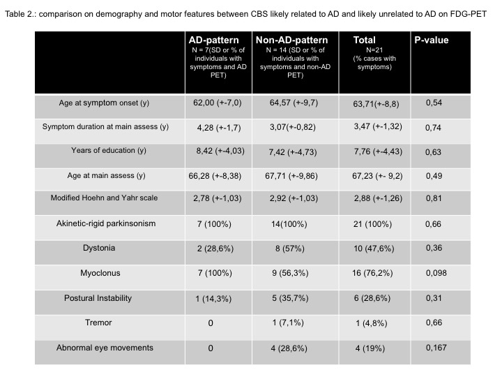
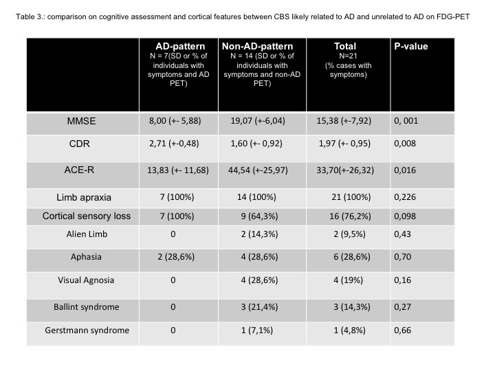
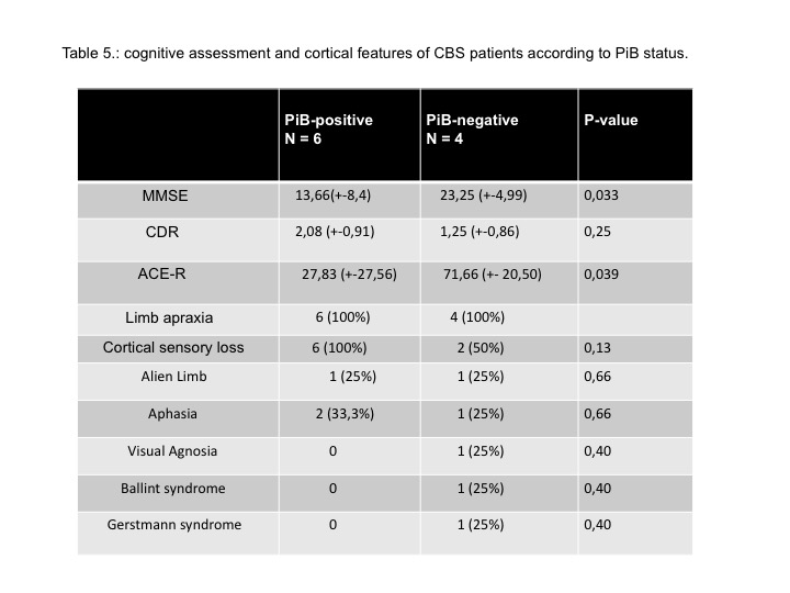
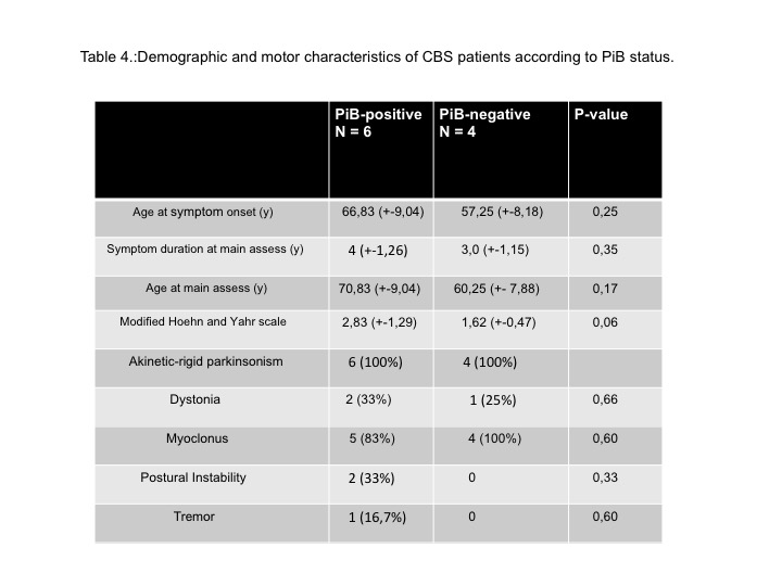
Results: Demography is shown in table 1. Parkinsonism was present in 100%, myoclonus in 76,2%, dystonia in 47,6%. Apraxia was the most prevalent cognitive feature. Cortical sensory loss was present in 76,2%, only two patients had alien limb phenomena. Fourteen patients had a non-AD FDG-PET pattern, and 7 had AD-pattern [figures 1, 2]. Regarding amyloid PET, six were PiB-positive and 4 PiB-negative [figures 3, 4]. Two patients with non-AD FDG-PET pattern in a total of 6 were PiB+. All 4 cases classified as having an AD-pattern were PiB+. All 4 cases of PiB- were classified previously as non-AD-pattern. FDG-PET subgroups showed no significant differences in age, gender, symptom duration and motor features [tables 2, 4]. There were significant differences on FDG-PET subgroups on MMSE score, Clinical Dementia Rating (CDR) scale, and Addenbrooke’s Cognitive Examination-Revised (ACE-R) [table3]. We also observed significant differences in PiB subgroups on MMSE and ACE-R [table5].
Conclusion: All patients with an AD pattern on FDG-PET were PiB+ and had lower ACE-R and MMSE scores despite age, gender, symptom duration, and motor features, suggesting more functional decline related to AD pathology. Functional molecular imaging might be a potential factor to predict CBS variants depicting their specific patterns.
References: 1. Armstrong MJ, Litvan I, Lang AE, Bak TH, Bhatia KP, Borroni B, et al. Criteria for the diagnosis of corticobasal degeneration.Neurology 2013;80:496–503. 2. Burrell JR, Hornberger M, Villemagne VL, Rowe CC, Hodges JR. Clinical profile of PiB-positive corticobasal syndrome. PLoS One. 2013;8:e61025. 3. Sha et al. Predicting amyloid status in corticobasal syndrome using modified clinical criteria, magnetic resonance imaging and fluorodeoxyglucose positron emission tomography.Alzheimer’s Research & Therapy . 2015 Mar 2;7(1):8.
To cite this abstract in AMA style:
J. Parmera, A. Coutinho, A. Neto, M. Aranha, C. Ono, C. Buchpiguel, R. Nitrini, E. Barbosa, S. Brucki. Deciphering Corticobasal Syndrome: are clinical features and FDG-PET imaging capable of predicting amiloyd status? [abstract]. Mov Disord. 2019; 34 (suppl 2). https://www.mdsabstracts.org/abstract/deciphering-corticobasal-syndrome-are-clinical-features-and-fdg-pet-imaging-capable-of-predicting-amiloyd-status/. Accessed April 22, 2025.« Back to 2019 International Congress
MDS Abstracts - https://www.mdsabstracts.org/abstract/deciphering-corticobasal-syndrome-are-clinical-features-and-fdg-pet-imaging-capable-of-predicting-amiloyd-status/

