Category: Parkinson's Disease: Neuroimaging
Objective: To investigate cortical mean diffusivity (cMD) as a sensitive measure to identify subtle microstructural cortical changes in Parkinson’s disease (PD) patients with probable REM sleep behaviour disorder (pRBD).
Background: REM sleep behaviour disorder (RBD) has been described as a symptom of PD associated with greater cognitive impairment [1] and brain atrophy [2][3]. Nevertheless, evidence regarding cortical atrophy patterns in PD pRBD patients is still scarce. Cortical MD has demonstrated high sensitivity to detect cortical microstructural damage in different neurodegenerative diseases, so it has been proposed as a novel imaging biomarker [4]. However, it has not been comprehensively investigated in PD.
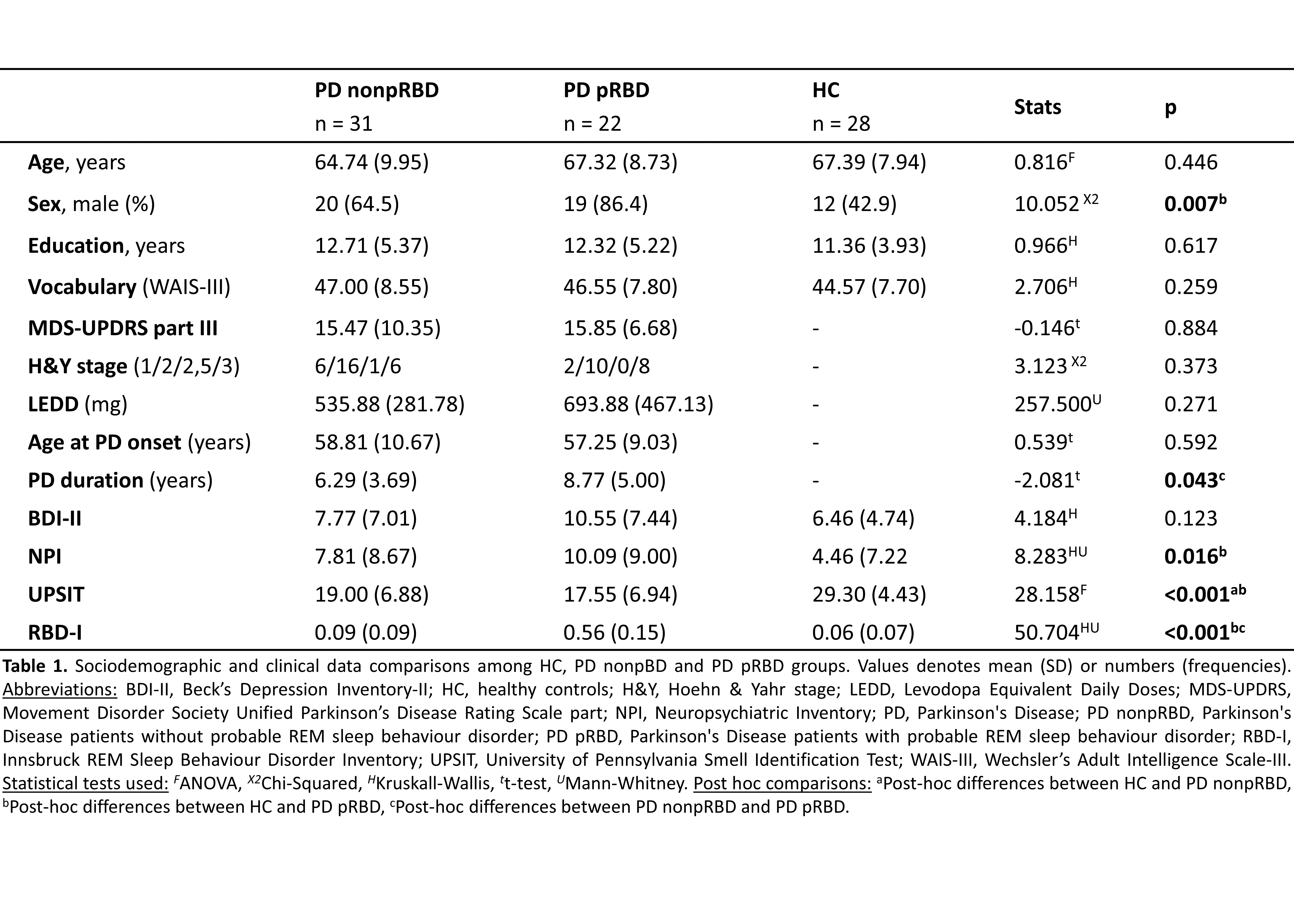
Method: Fifty-three PD patients, classified as PD pRBD (n=22) and PD nonpRBD (n=31) through the Innsbruck RBD Inventory, along with 28 healthy controls (HC) were assessed using a neuropsychological testing and 3-dimensional T1-weighted, as well as diffusion-weighted MRI on a 3T scanner. Cortical MD and cortical thickness (CTh) were used as surface-based approaches to obtain measures of cortical brain changes. Two-class group comparisons of a general linear model were performed (p value set at 0.05), considering sex and PD evolution as confounding variables when it was required [table1]. Cohen’s d effect size for both approaches was computed.
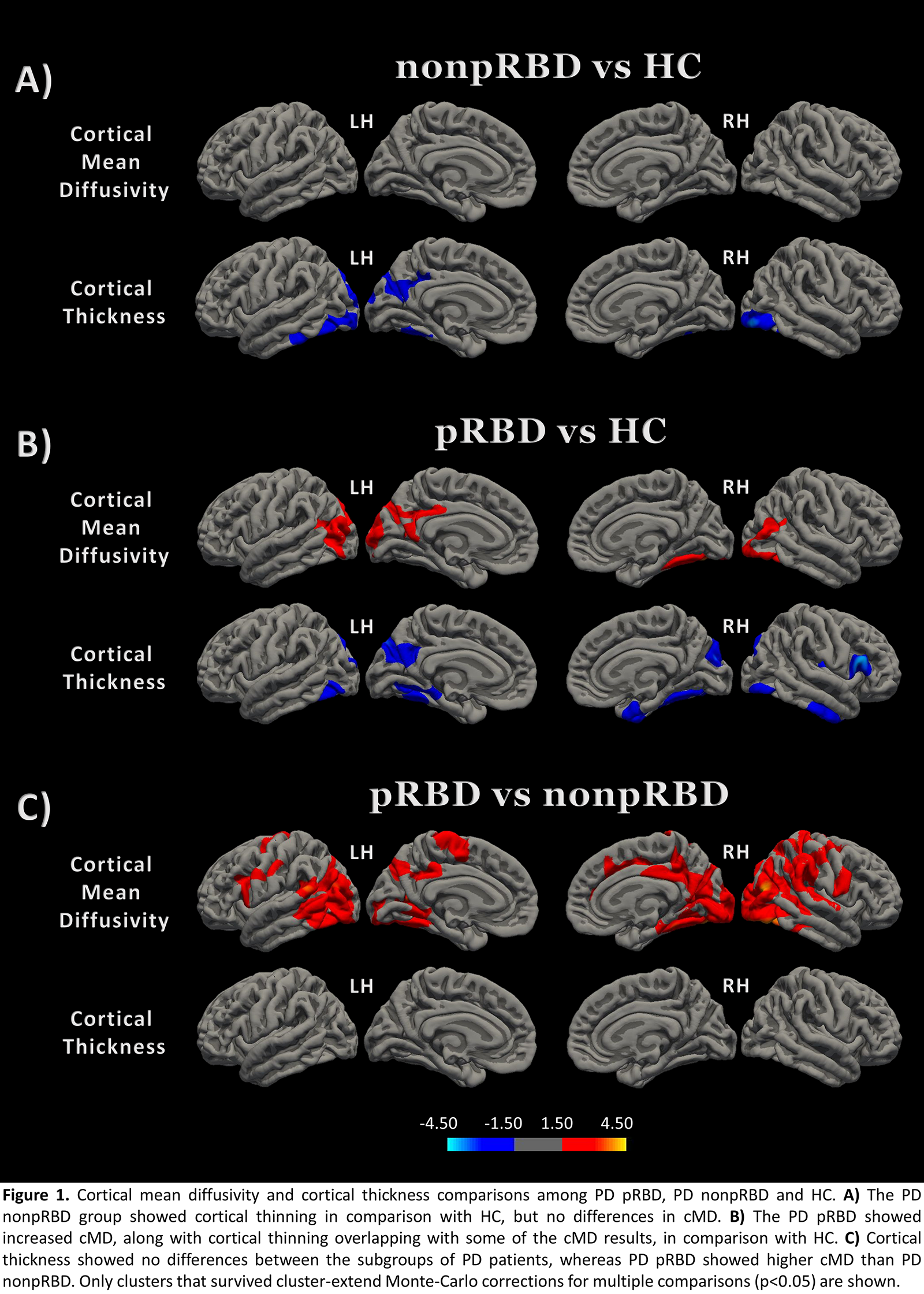
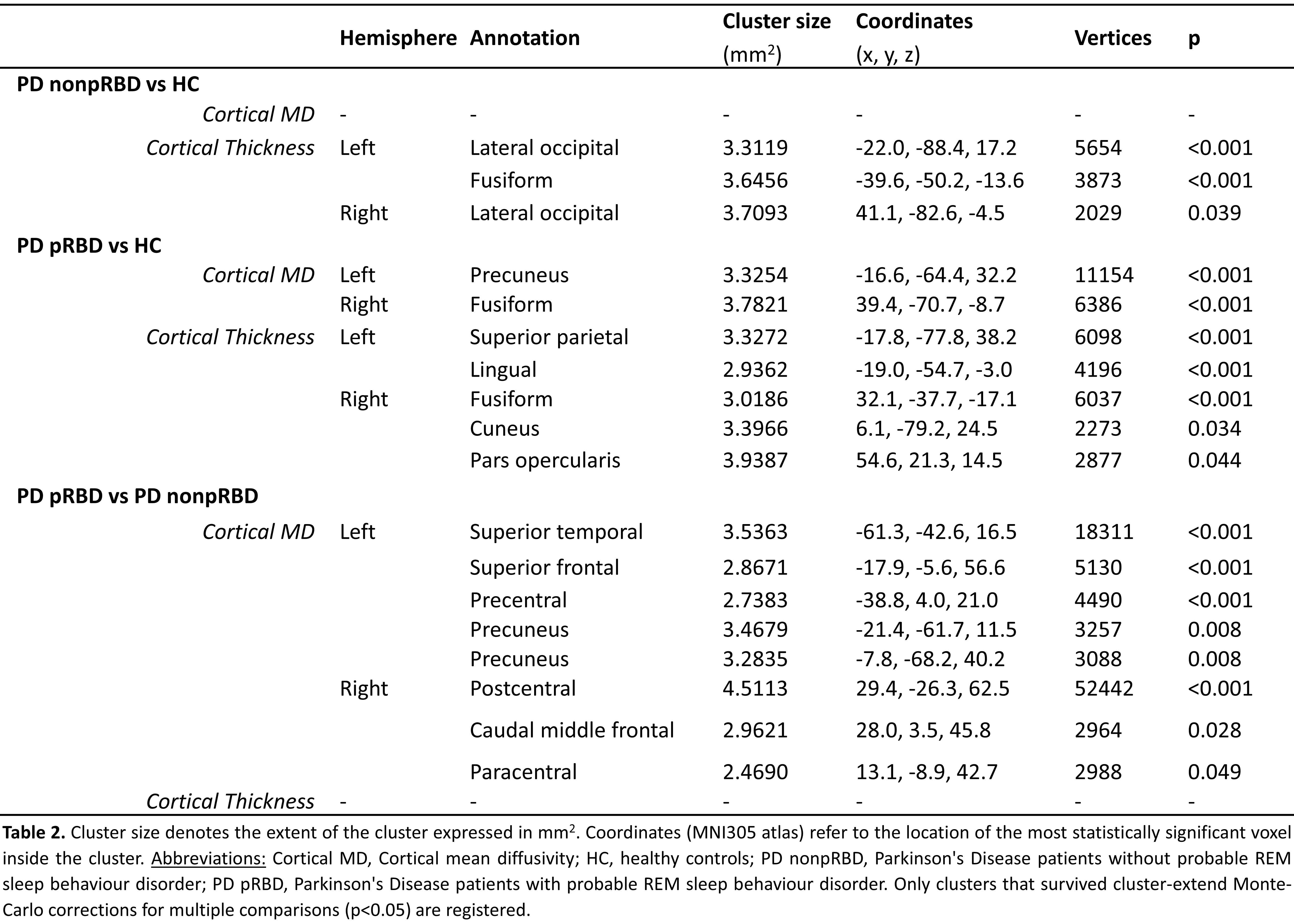
Results: PD pRBD patients showed higher cMD than PD nonpRBD in the left superior temporal, superior frontal, and precentral gyri, precuneus cortex, as well as in the right middle frontal and postcentral gyri and paracentral lobule with effect sizes between 0.5 and 1.06, while CTh contrast showed no differences. PD pRBD patients also showed increased bilateral posterior cMD in comparison with HC with effect sizes between 0.5 and 1.11. These results partially overlapped with CTh results that showed effect sizes between 0.5 and 0.98. PD nonpRBD patients showed no differences in cMD when compared with HC but showed cortical thinning in the left fusiform gyrus and lateral occipital cortex bilaterally with moderate effect sizes between 0.5 and 0.68 [figure1][table2].
Conclusion: Our results have unveiled a posterior pattern of microstructural damage in the PD pRBD group. Cortical MD has shown more sensitivity than CTh displaying significant microstructural differences between the PD subgroups, indicating the utility of this imaging biomarker to study early cortical damage in PD.
References: [1] Trout J, Christiansen T, Bulkley MB, et al. Cognitive Impairments and Self-Reported Sleep in Early-Stage Parkinson’s Disease with Versus without Probable REM Sleep Behavior Disorder. Brain Sci. 2019;10(1).
[2] Boucetta S, Salimi A, Dadar M, et al. Structural Brain Alterations Associated with Rapid Eye Movement Sleep Behavior Disorder in Parkinson’s Disease. Sci Rep. 2016;6:26782.
[3] Oltra J, Uribe C, Segura B, et al. Brain atrophy pattern in de novo Parkinson’s disease with probable RBD associated with cognitive impairment. npj Park Dis. 2022;24(8):60.
[4] Illán-Gala I, Montal V, Pegueroles J, et al. Cortical microstructure in the amyotrophic lateral sclerosis–frontotemporal dementia continuum. Neurology. 2020;95:e2565–76.
To cite this abstract in AMA style:
J. Pardo, V. Montal, A. Campabadal, J. Oltra, C. Uribe, I. Roura, MJ. Martí, Y. Compta, A. Iranzo, J. Fortea, C. Junqué, B. Segura. Cortical mean diffusivity changes in Parkinson’s disease with probable RBD [abstract]. Mov Disord. 2023; 38 (suppl 1). https://www.mdsabstracts.org/abstract/cortical-mean-diffusivity-changes-in-parkinsons-disease-with-probable-rbd/. Accessed December 18, 2025.« Back to 2023 International Congress
MDS Abstracts - https://www.mdsabstracts.org/abstract/cortical-mean-diffusivity-changes-in-parkinsons-disease-with-probable-rbd/


