Category: Neuroimaging (Non-PD)
Objective: To correlate nigrosome-1 (NG1) imaging and SPECT 123I-Ioflopane findings to determine the feasibility and diagnostic value of NG1 imaging in a real-world clinical setting.
Background: Since the description of NG1 as the dopaminergic neuron rich area in the dorsolateral border of substantia nigra compacta (SNc) that is most affected in Parkinson’s disease[1], ongoing research has aimed to describe the radiological characteristics of NG1. However, non-standardized methods limit the use of NG1 imaging in clinical practice and studies correlating results between NG1 imaging and standard nuclear medicine are sparse[2][3].
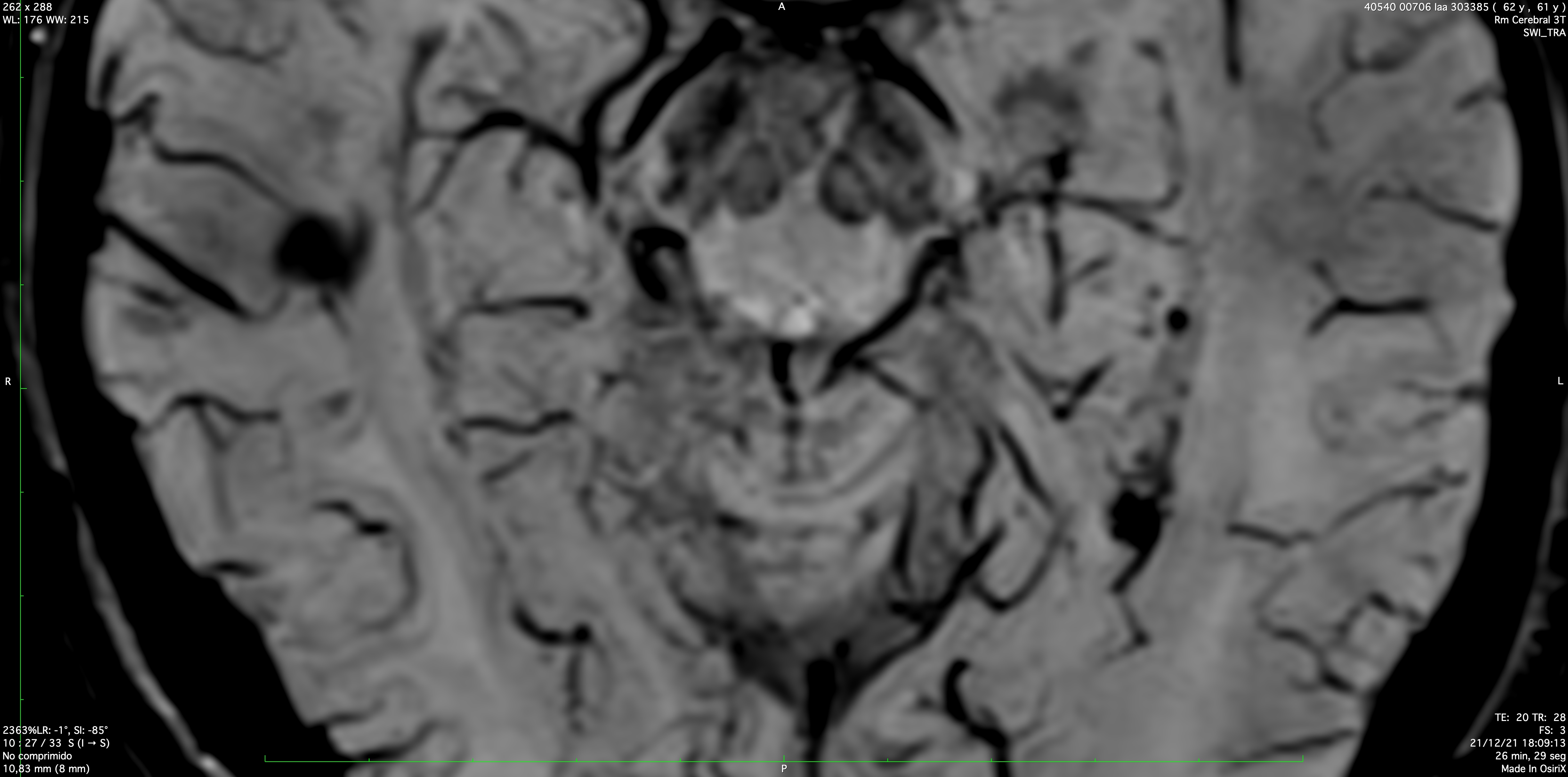
Method: We performed a chart review of patients with (<5 year) parkinsonism seen in our movement disorders unit and studied with both 3 Tesla MRI including high-resolution susceptibility-weighted imaging (SWI) to detect NG1 and SPECT 123I-Ioflupane within 6 months of each other. Diagnostic tests were categorically classified by neuroradiology and nuclear medicine experts as negative (normal), unilaterally positive or bilaterally positive (each rater blinded to other’s rating). MRI was deemed positive if SNc dorsolateral hyperintensity or swallow-tail sign was absent [4]. Concordance analysis for imaging techniques was performed with Cohen’s kappa test.
Results: Ten patients (7 males), mean age (+SD) 62.5(+12.6) years and median disease duration of 2 years (range 0.5-5) were analyzed. Three patients with slight parkinsonism, associated with dystonic tremor and one with parkinsonism associated with frontal ataxia had normal SPECT and NG1 imaging. Four patients with bilateral asymmetric parkinsonism had positive bilateral SPECT and NG1 imaging. Two patients who also had bilateral asymmetric parkinsonism displayed disparate results: one had bilateral positive SPECT and only left positive NG1 [figure 1], while the other had unilaterally positive SPECT with normal NG1 imaging. Cohen’s kappa test showed substantial agreement per patient (k=0.75) and per nigrosome (k=0.81).
Conclusion: In our limited experience, qualitative analysis of NG1 has reasonably good concordance with SPECT 123I-Ioflupane results in patients with parkinsonism. This preliminary data suggests that NG1 imaging may be a useful diagnostic tool in our clinical setting. However, larger prospective studies are warranted to this.
References: 1. Damier P, Hirsch EC, Agid Y, Graybiel AM. The substantia nigra of the human brain. II. Patterns of loss of dopamine-containing neurons in Parkinson’s disease. Brain. 1999 Aug;122 (Pt 8):1437-48.
2. Sung YH, Lee J, Nam Y, Shin HG, Noh Y,Hwang KH, et al. Initial diagnostic work up of parkinsonism: Dopamine transporter positron emission tomography versus susceptibility map weighted imaging at 3T.Parkinsonism Relat Disord. 2019 May;62:171-178.
3. Chau MT, Todd G, Wilcox R, Agzarian M, Bezak E. Diagnostic accuracy of the appearance of Nigrosome-1 on magnetic resonance imaging in Parkinson’s disease: A systematic review and meta-analysis. Parkinsonism Relat Disord. 2020 Sep;78:12-20.
4. Arribarat G, De Barros A, Péran P. Modern Brainstem MRI Techniques for the Diagnosis of Parkinson’s Disease and Parkinsonisms. Front Neurol. 2020 Aug 4;11:791.
To cite this abstract in AMA style:
A. López-Jiménez, J. álvarez-Linera, JF. Boan, I. Pareés, M. Kurtis. Correlation between 3 Tesla Brain MRI Nigrosome-1 imaging and SPECT 123I-Ioflupane in parkinsonian disorders [abstract]. Mov Disord. 2022; 37 (suppl 2). https://www.mdsabstracts.org/abstract/correlation-between-3-tesla-brain-mri-nigrosome-1-imaging-and-spect-123i-ioflupane-in-parkinsonian-disorders/. Accessed December 30, 2025.« Back to 2022 International Congress
MDS Abstracts - https://www.mdsabstracts.org/abstract/correlation-between-3-tesla-brain-mri-nigrosome-1-imaging-and-spect-123i-ioflupane-in-parkinsonian-disorders/

