Session Information
Date: Thursday, June 8, 2017
Session Title: Parkinson's Disease: Neuroimaging And Neurophysiology
Session Time: 1:15pm-2:45pm
Location: Exhibit Hall C
Objective: To investigate correlations between the sub-regional dopamine release (DR) in the striatum of Parkinson’s disease (PD) subjects and sub-components of motor performance evaluated according to UPDRS III.
Background: Previous PET studies of DR in PD used either simple geometric or anatomy-defined regions of interest (ROIs) for each sub-structure in the striatum. It is however also of interest to investigate whether specific PD motor symptoms may be related to a somatotopic degeneration in distinct sub-regions of the putamen, caudate and ventral striatum (VS).
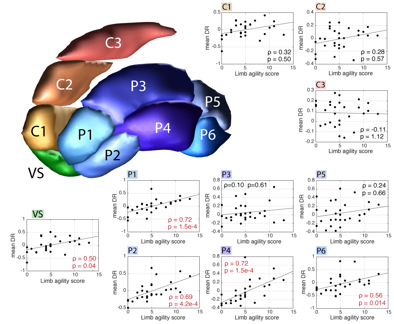
Methods: DR images for 14 mild- to moderately-impaired PD subjects were calculated as the difference between the baseline and post-stimulus (rTMS) parametric PET images of the [11C]raclopride (RAC) non-displaceable binding potential (BPND) obtained with SRTM2. The DR images were co-registered with acquired MRI images that were segmented. Segmentations of the putamen, caudate, and VS were warped to a common template; the same transformations were applied to the DR images. The template was subdivided into 10 sub-regions [figure1] (Fig. 1). The mean sub-regional DR was regressed (Spearman’s ρ) against the contralateral UPDRS III sub-scores: limb agility, bradykinesia, rigidity, and facial scores.
Results: After correction for multiple comparisons, limb agility was significantly (p<10-3) correlated with DR in VS and in the anterior-inferior putamen sub-regions (P1, P2, P4 and P6) (Fig. 1); the was no significant correlation in the caudate. With bradykinesia, significant correlations were found in the sub-regions P1 (ρ=0.54, p=0.02), P2 (ρ=0.61, p=0.005) and P4 (ρ=0.60, p=0.007). Rigidity and facial scores were not correlated with DR anywhere in the striatum. Voxel-wise correlation analysis revealed regions in the striatum with highest ρ values for correlations of DR versus limb agility and bradykinesia [figure2] (Fig 2).
Conclusions: Results demonstrate that rTMS-induced DR in the putamen is correlated with specific aspects of motor impairment in PD; the degree of correlation varies across sub-regions in the putamen. This is indicative of a specific connectivity that may exist between motor circuits and the sub-regions; in future work this connectivity will be further investigated using high-resolution PET imaging, to conduct a correlation analysis between cognitive function and DR in the caudate.
To cite this abstract in AMA style:
I. Klyuzhin, M. Sacheli, N. Vafai, E. Shahinfard, B. Lakhani, J. Neva, J. Fu, J. McKenzie, N. Neilson, K. Dinelle, L. Boyd, A. Stoessl, V. Sossi. Correlation analysis between dopamine release in striatal sub-regions and motor impairment in Parkinson’s disease subjects [abstract]. Mov Disord. 2017; 32 (suppl 2). https://www.mdsabstracts.org/abstract/correlation-analysis-between-dopamine-release-in-striatal-sub-regions-and-motor-impairment-in-parkinsons-disease-subjects/. Accessed December 24, 2025.« Back to 2017 International Congress
MDS Abstracts - https://www.mdsabstracts.org/abstract/correlation-analysis-between-dopamine-release-in-striatal-sub-regions-and-motor-impairment-in-parkinsons-disease-subjects/


