Category: Neuroimaging (Non-PD)
Objective: To investigate the dopaminergic neural tracts of healthy individuals using Diffusion MRI on Human Connectome Project Data.
Background: Classically, the DA system has been described with respect to three distinct pathways, described here as the conventional pathway heuristic. In the nigrostriatal pathway, DA neurons from the substantia nigra pars compacta (SNc) project to the dorsal striatum (DS). In the mesolimbic pathway, ventral tegmental area (VTA) dopaminergic neurons project to the ventral striatum (VS). In the mesocortical pathway, VTA dopaminergic neurons project to the prefrontal cortex (PFC).
Although providing a convenient explanation for DA-mediated behaviours, the conventional pathway heuristic is now thought to be an oversimplification of SNc/VTA dopaminergic circuits.
Method: 3T MRI Data from the WU-Minn 1200 subjects Release (S1200) of March 01, 2017 were utilized in this study. FSL PROBTRACKX was used to calculate connectivity density from seed voxels in the SNc and VTA to striatal and cortical targets. 5000 streamlines were seeded from each seed voxel in the SNc and VTA to proximal probability density functions previously established by FSL BEDPOST. Streamlines that made contact with striatal or cortical subregions were tallied. Connectivity was defined as the proportion of streamlines that made contact with a target region.
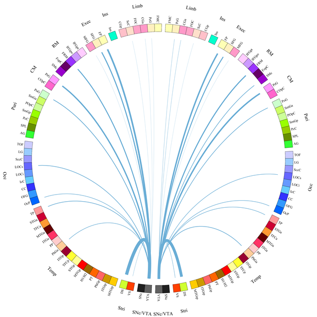
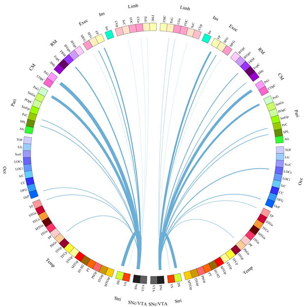
Results: We measured the following findings that are at odds with the conventional pathway heuristic (Fig. 1 &2). A) The SNc (M = 11.169%, SEM = 0.359) and VTA (M = 11.189%, SEM = 0.342) had statistically equivalent (F(1,98) <1) connectivity densities to and from the dorsal striatum. B) The ventral striatum had greater connectivity density to and from the SNc (M = 0.120%, SEM = 0.010) than to and from the VTA(M = 0.063%, SEM = 0.006; F(1,98) = 78.850, MSe = 0.004, p < 0.001). C) The prefrontal cortex had greater connectivity density to and from the SNc (M = 0.086%, SEM = 0.005) than to and from the VTA (M = 0.043%, SEM = 0.003; F(1,98) = 142.676, MSe = 0.008, p < 0.001).
Conclusion: Our results add evidence for neural pathways between the VTA–dorsal striatum, SNc–ventral striatum, and SNc–prefrontal cortex.
To cite this abstract in AMA style:
N. Handfield-Jones, A. Owen, A. Khan, P. Macdonald. Connectomic Analysis of SNc and VTA Projections to the Striatum and Cortex [abstract]. Mov Disord. 2021; 36 (suppl 1). https://www.mdsabstracts.org/abstract/connectomic-analysis-of-snc-and-vta-projections-to-the-striatum-and-cortex/. Accessed April 26, 2025.« Back to MDS Virtual Congress 2021
MDS Abstracts - https://www.mdsabstracts.org/abstract/connectomic-analysis-of-snc-and-vta-projections-to-the-striatum-and-cortex/


