Category: Parkinson's Disease: Neuroimaging
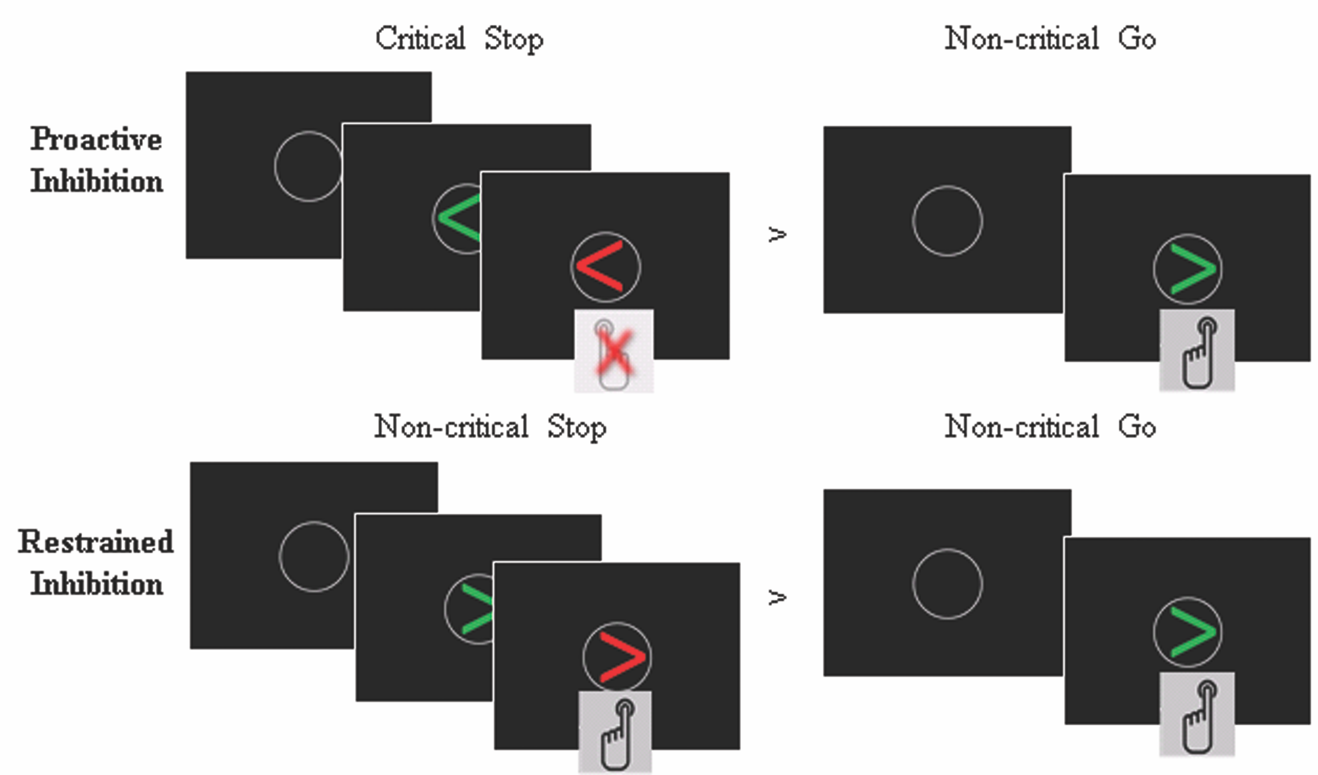
Objective: To investigate response inhibition in Parkinson’s disease (PD) patients with impulse control disorders. Specifically, the neural underpinnings of two aspects of inhibition: proactive inhibition –inhibition that has been prepared beforehand– and restrained inhibition –inhibition of an impulse to inhibit.
Background: Impulse control disorders have a high prevalence in PD and have a strong negative impact on the quality of life of those affected. Despite impulsivity having classically been associated with response inhibition deficits, previous evidence from PD patients with impulse control disorder does not show behavioral dysfunction in response inhibition, suggesting the presence of compensatory mechanisms.
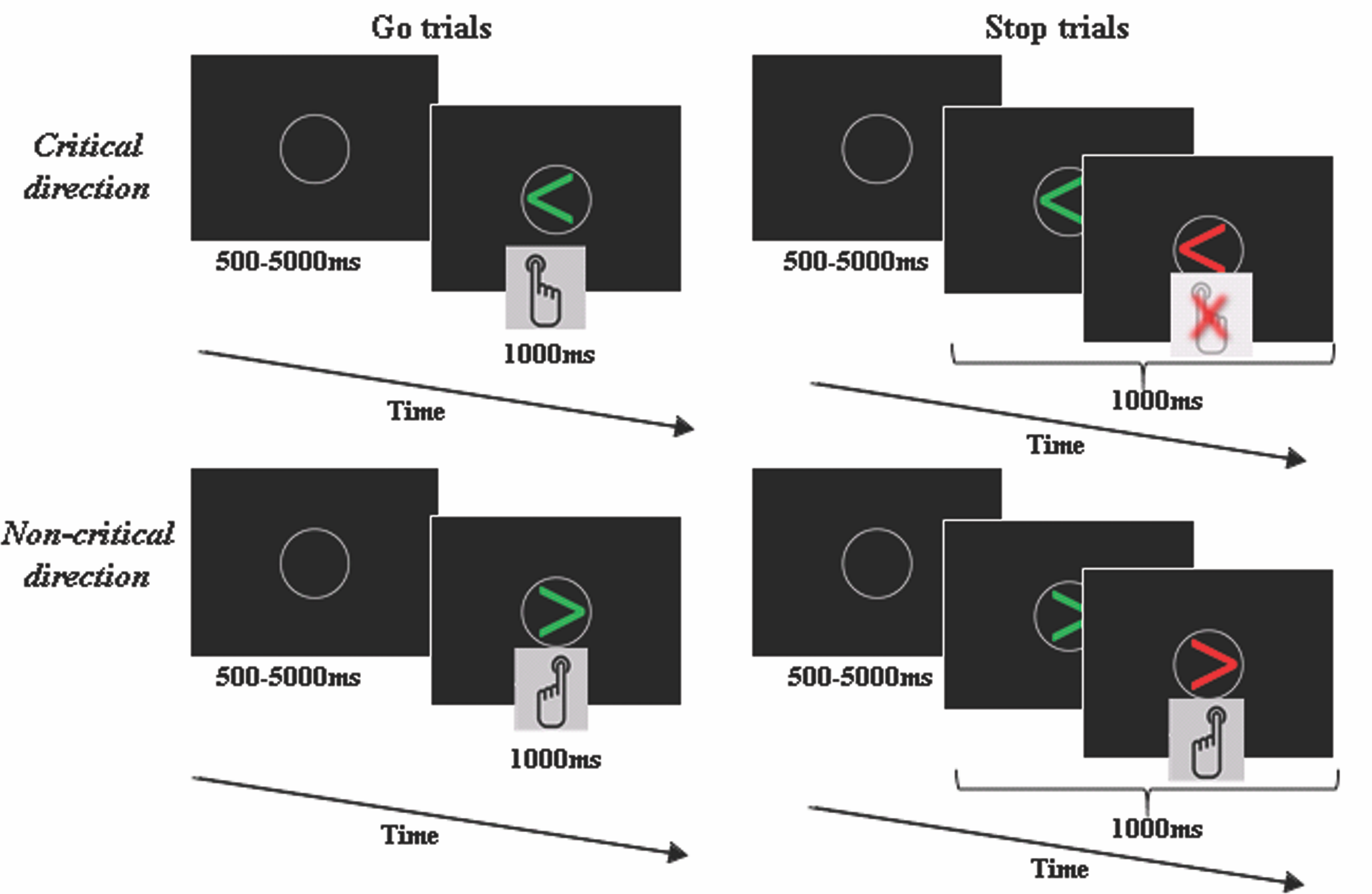
Method: Eighteen PD patients with impulse control disorders, 17 PD patients without the disorder, and 15 healthy controls performed a version of the conditional Stop Signal Task during functional magnetic resonance imaging. We conducted whole-brain contrasts, regions of interest, and whole-brain functional connectivity analyses.
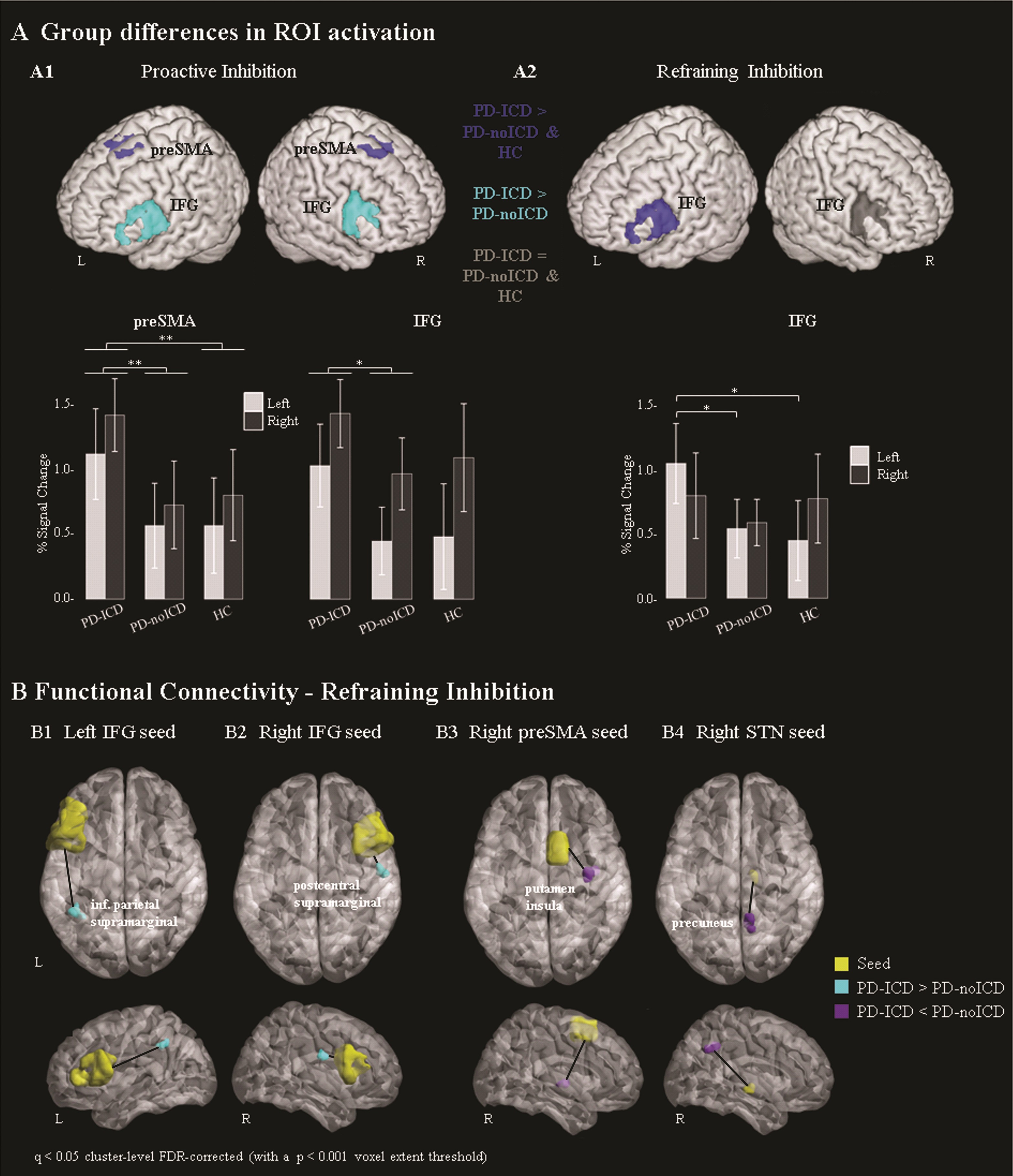
Results: PD patients with impulse control disorder exhibited bilateral hyperactivation of two areas of the stopping network – inferior frontal gyrus (IFG) and presupplementary motor area – while performing proactive inhibition. When engaged in restrained inhibition, they showed hyperactivation of the left IFG, an area linked to action monitoring. Additionally, restrained inhibition showed changes in the functional coactivation of inhibitory regions with left inferior parietal cortex and the right supramarginal gyrus, as well as reduced coactivation between the right subthalamic nucleus and the precuneus.
Conclusion: PD patients with impulse control disorder completed the inhibition task correctly, while showing altered engagement of inhibitory and attentional areas. During proactive inhibition they showed bilateral hyperactivation of two inhibitory regions, while during restrained inhibition they showed additional involvement of attentional areas responsible for alerting and orienting, suggesting that depending on the type of inhibition, PD patients with impulse control disorder deploy additional mechanisms to maintain an adequate inhibitory performance.
This work was presented at the SfN Global Connectome Virtual Event in January, 2021.
To cite this abstract in AMA style:
T. Esteban-Peñalba, P. Paz-Alonso, I. Navalpotro-Gomez, M. Rodriguez-Oroz. Compensatory functional mechanisms of response inhibition in Parkinson’s Disease with impulse control disorders [abstract]. Mov Disord. 2021; 36 (suppl 1). https://www.mdsabstracts.org/abstract/compensatory-functional-mechanisms-of-response-inhibition-in-parkinsons-disease-with-impulse-control-disorders/. Accessed April 26, 2025.« Back to MDS Virtual Congress 2021
MDS Abstracts - https://www.mdsabstracts.org/abstract/compensatory-functional-mechanisms-of-response-inhibition-in-parkinsons-disease-with-impulse-control-disorders/


