Category: Parkinson's Disease: Neuroimaging
Objective: We sought to determine whether functional network abnormalities seen in patients with Parkinson’s disease (PD) and cognitive decline reflect the loss of the default mode network (DMN), the gain of an abnormal network topography, or a combination of both.
Background: Cognitive decline, a common symptom in patients with PD, has been associated with stereotyped changes in normal resting state networks and cognition related networks evolving over time [1]. It is not known whether the PD cognition-related pattern (PDCP) represents the loss of the DMN as a manifestation of the disease process.
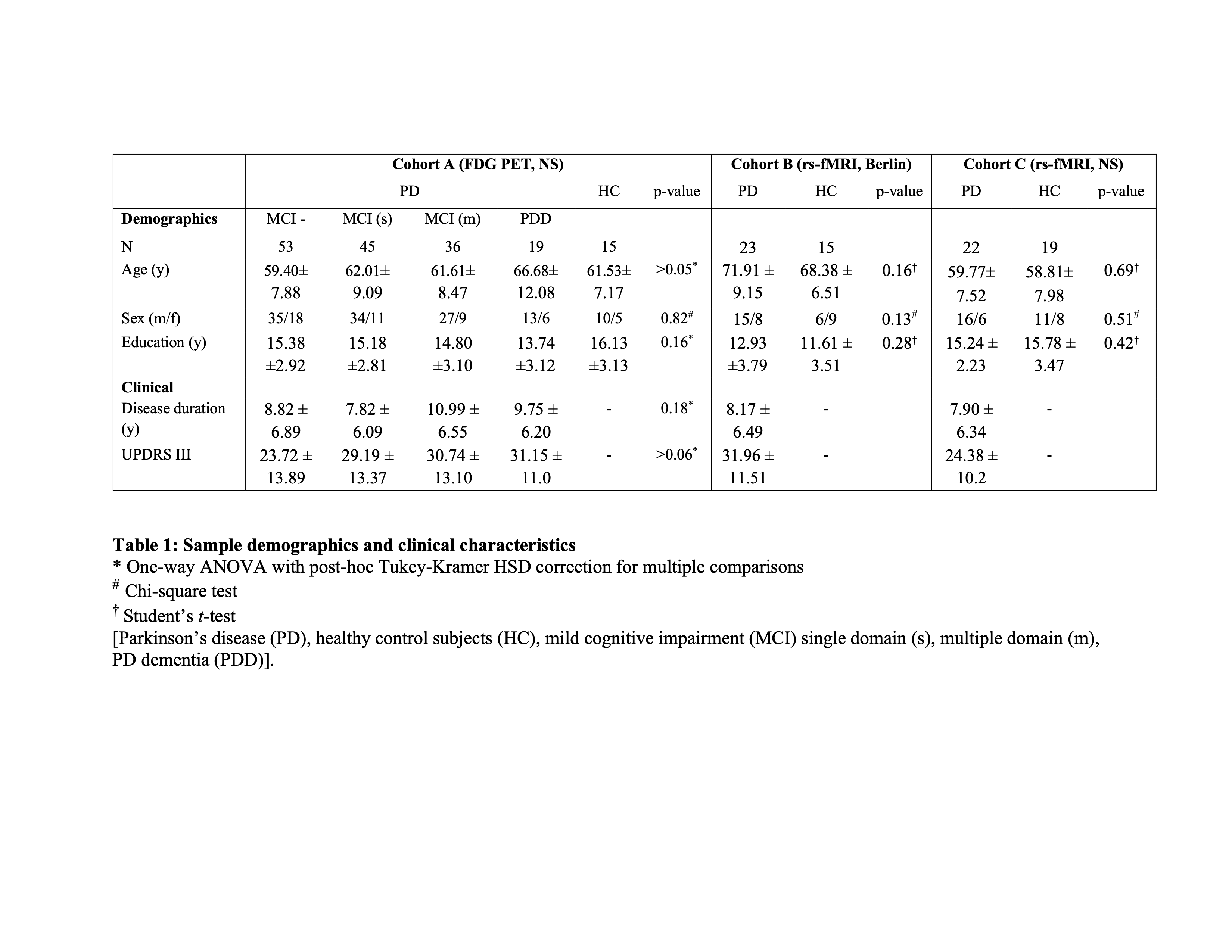
Method: We first analyzed 18F-FDG-PET images to evaluate network expression levels in a large PD sample with varying cognitive performance at NorthShore (NS) [table 1]. We then used independent component analysis to identify fPDCP and DMN topographies in resting-state fMRI (rs-fMRI) data from an independent population in Berlin [table 1]. The spatial relationship of the two topographies was determined at the subnetwork level. The topographies were further compared to previously described PDCP networks from NS to evaluate their consistency across cohorts, imaging platforms, and medication state.
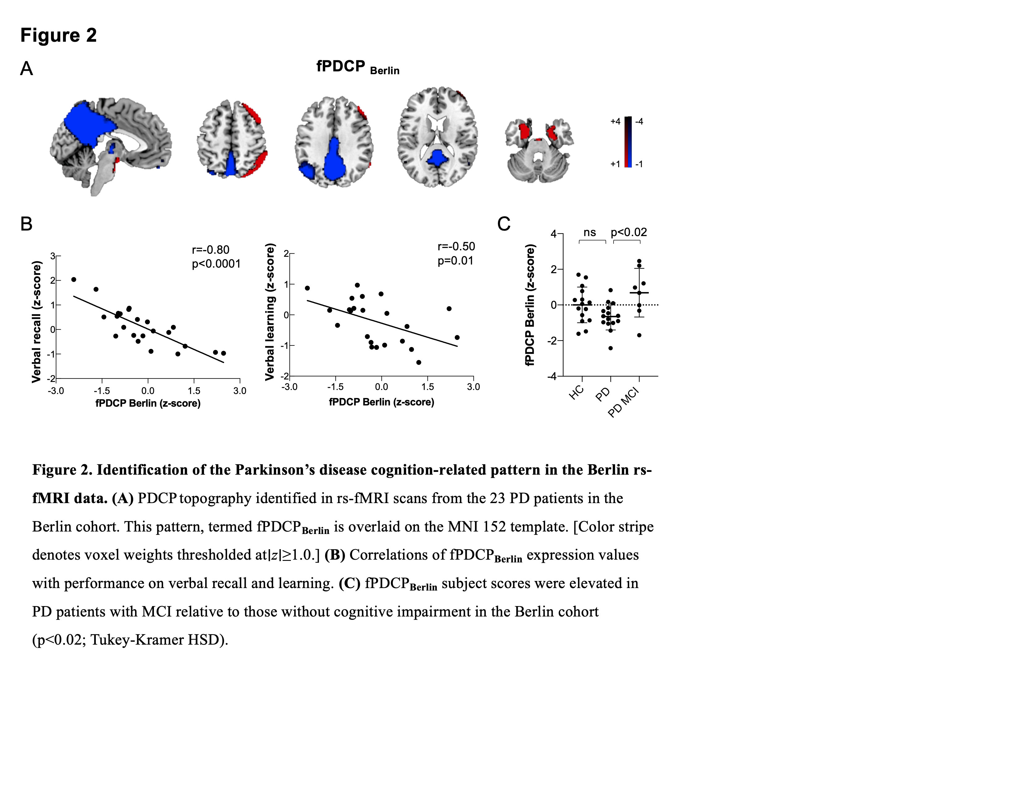
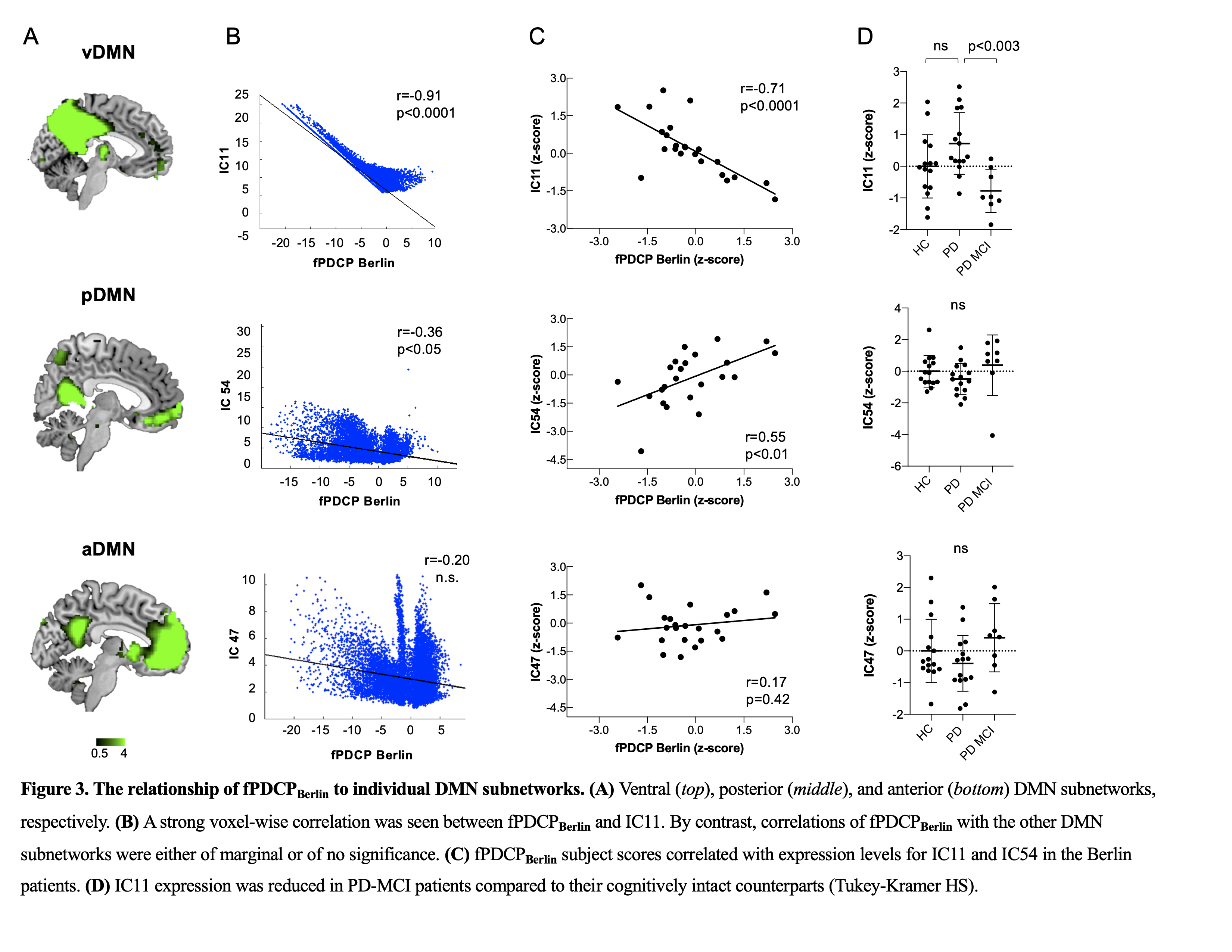
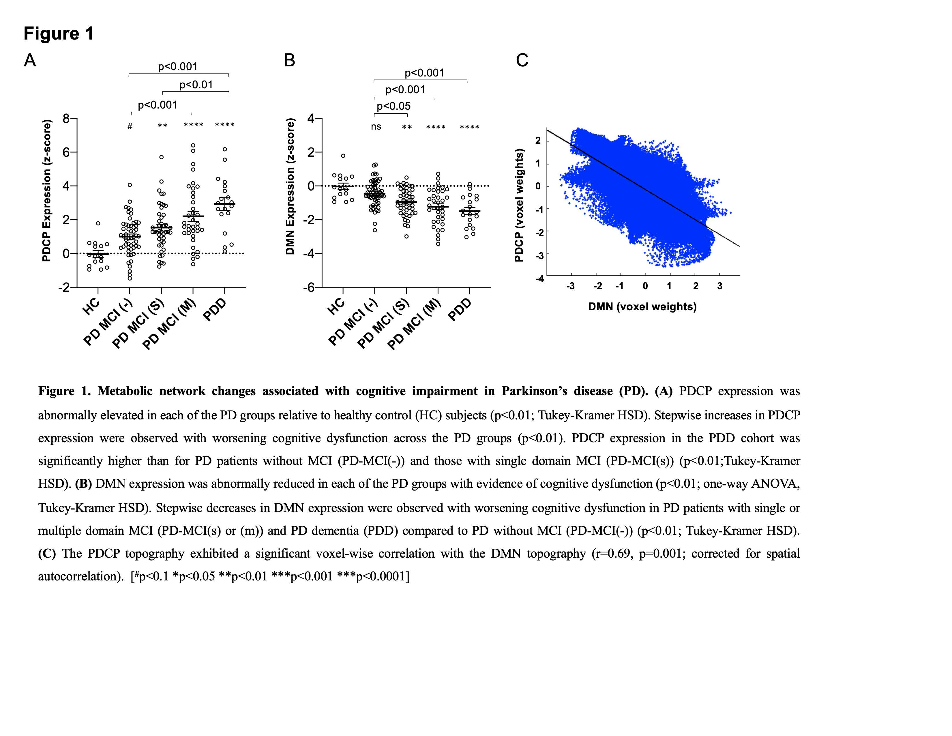
Results: Cognitive impairment in PD is associated with increases in metabolic PDCP expression and reflects partial loss of DMN activity (figure 1). We identified a fPDCP topography in an independent cohort scanned with rs-fMRI in the on-medication state (figure 2). fPDCPBERLIN expression correlated with verbal memory performance and was elevated in PD patients with MCI compared to their counterparts without cognitive deficits. This topography resembled the previously described PDCPNS patterns from FDG-PET and rs-fMRI data scanned in the off-medication state. The rs-MRI analysis uncovered that PDCP selectively involves the ventral DMN subnetwork (precuneus and PCC), while the anterior and posterior subcircuits of the DMN are not affected (figure 3). Importantly, the PDCP goes beyond the DMN topography with activity changes in dorsolateral prefrontal and medio-temporal areas that are unrelated to the DMN space.
Conclusion: The findings show that the PDCP is a reproducible cognition-related network that is topographically distinct from the DMN. The utility of the PDCP as a network biomarker of cognitive dysfunction in PD is further highlighted by its independence of imaging platforms and treatment condition.
References: Schindlbeck, K. A., & Eidelberg, D. (2018). Network imaging biomarkers: insights and clinical applications in Parkinson’s disease. The Lancet Neurology, 17(7), 629–640.
To cite this abstract in AMA style:
K. Schindlbeck, A. Vo, P. Mattis, K. Villringer, F. Marzinzik, J. Fiebach, D. Eidelberg. Cognition-related functional topographies in Parkinson’s disease: Localized loss of the ventral default mode network [abstract]. Mov Disord. 2021; 36 (suppl 1). https://www.mdsabstracts.org/abstract/cognition-related-functional-topographies-in-parkinsons-disease-localized-loss-of-the-ventral-default-mode-network/. Accessed April 21, 2025.« Back to MDS Virtual Congress 2021
MDS Abstracts - https://www.mdsabstracts.org/abstract/cognition-related-functional-topographies-in-parkinsons-disease-localized-loss-of-the-ventral-default-mode-network/


